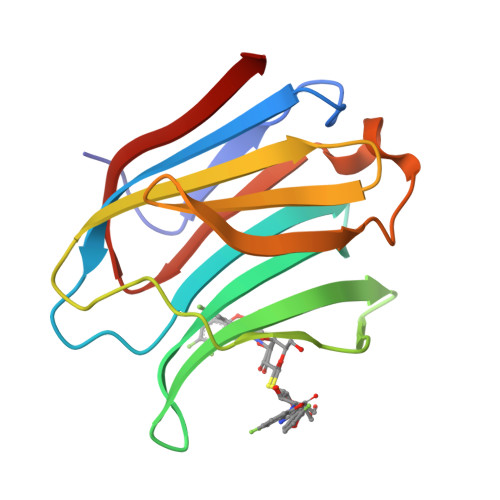
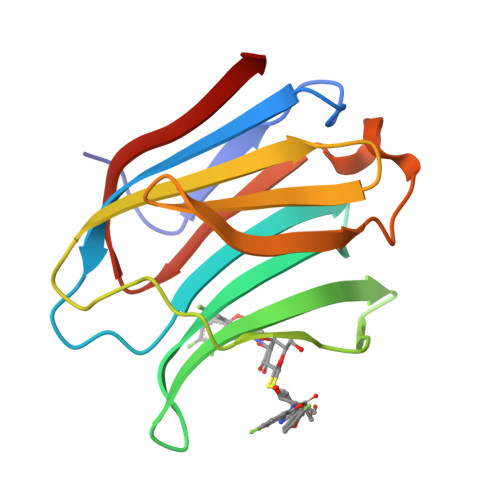
5OAX

Galectin-3c in complex with thiogalactoside derivate


Experimental Data Snapshot

  • Method: X-RAY DIFFRACTION
  • Resolution: 1.20 Å
  • R-Value Free: 0.132 
  • R-Value Work: 0.103 
  • R-Value Observed: 0.104 

wwPDB Validation   3D Report Full Report


Ligand Structure Quality Assessment 


This is version 1.2 of the entry. See complete history


Literature

Systematic Tuning of Fluoro-galectin-3 Interactions Provides Thiodigalactoside Derivatives with Single-Digit nM Affinity and High Selectivity.

Peterson, K.Kumar, R.Stenstrom, O.Verma, P.Verma, P.R.Hakansson, M.Kahl-Knutsson, B.Zetterberg, F.Leffler, H.Akke, M.Logan, D.T.Nilsson, U.J.

(2018) J Med Chem 61: 1164-1175

  • DOI: https://doi.org/10.1021/acs.jmedchem.7b01626
  • Primary Citation of Related Structures:  
    5OAX, 5ODY

  • PubMed Abstract: 

    Symmetrical and asymmetrical fluorinated phenyltriazolyl-thiodigalactoside derivatives have been synthesized and evaluated as inhibitors of galectin-1 and galectin-3. Systematic tuning of the phenyltriazolyl-thiodigalactosides' fluoro-interactions with galectin-3 led to the discovery of inhibitors with exceptional affinities (K d down to 1-2 nM) in symmetrically substituted thiodigalactosides as well as unsurpassed combination of high affinity (K d 7.5 nM) and selectivity (46-fold) over galectin-1 for asymmetrical thiodigalactosides by carrying one trifluorphenyltriazole and one coumaryl moiety. Studies of the inhibitor-galectin complexes with isothermal titration calorimetry and X-ray crystallography revealed the importance of fluoro-amide interaction for affinity and for selectivity. Finally, the high affinity of the discovered inhibitors required two competitive titration assay tools to be developed: a new high affinity fluorescent probe for competitive fluorescent polarization and a competitive ligand optimal for analyzing high affinity galectin-3 inhibitors with competitive isothermal titration calorimetry.


  • Organizational Affiliation

    Centre for Analysis and Synthesis, Department of Chemistry, Lund University , Box 124, SE-221 00 Lund, Sweden.


Macromolecules
Find similar proteins by:  (by identity cutoff)  |  3D Structure
Entity ID: 1
MoleculeChains Sequence LengthOrganismDetailsImage
Galectin-3138Homo sapiensMutation(s): 0 
Gene Names: LGALS3MAC2
UniProt & NIH Common Fund Data Resources
Find proteins for P17931 (Homo sapiens)
Explore P17931 
Go to UniProtKB:  P17931
PHAROS:  P17931
GTEx:  ENSG00000131981 
Entity Groups  
Sequence Clusters30% Identity50% Identity70% Identity90% Identity95% Identity100% Identity
UniProt GroupP17931
Sequence Annotations
Expand
  • Reference Sequence
Small Molecules
Ligands 1 Unique
IDChains Name / Formula / InChI Key2D Diagram3D Interactions
9Q5 (Subject of Investigation/LOI)
Query on 9Q5

Download Ideal Coordinates CCD File 
B [auth A]5,6-bis(fluoranyl)-3-[[(2~{S},3~{R},4~{S},5~{S},6~{R})-2-[(2~{S},3~{R},4~{S},5~{R},6~{R})-4-[4-(3-fluorophenyl)-1,2,3-triazol-1-yl]-6-(hydroxymethyl)-3,5-bis(oxidanyl)oxan-2-yl]sulfanyl-6-(hydroxymethyl)-3,5-bis(oxidanyl)oxan-4-yl]oxymethyl]chromen-2-one
C30 H30 F3 N3 O11 S
GCZYLGQPCXFNES-NFWFBJQFSA-N
Experimental Data & Validation

Experimental Data

  • Method: X-RAY DIFFRACTION
  • Resolution: 1.20 Å
  • R-Value Free: 0.132 
  • R-Value Work: 0.103 
  • R-Value Observed: 0.104 
  • Space Group: P 21 21 21
Unit Cell:
Length ( Å )Angle ( ˚ )
a = 36.19α = 90
b = 58.15β = 90
c = 62.81γ = 90
Software Package:
Software NamePurpose
REFMACrefinement

Structure Validation

View Full Validation Report


Ligand Structure Quality Assessment 


Entry History 

Deposition Data

Revision History  (Full details and data files)

  • Version 1.0: 2018-05-02
    Type: Initial release
  • Version 1.1: 2019-10-16
    Changes: Data collection
  • Version 1.2: 2024-05-08
    Changes: Data collection, Database references