5NNP | pdb_00005nnp

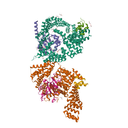
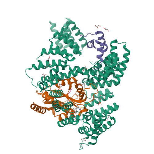
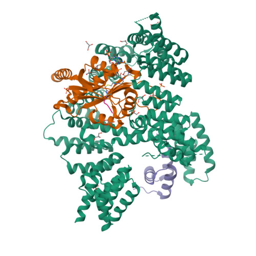
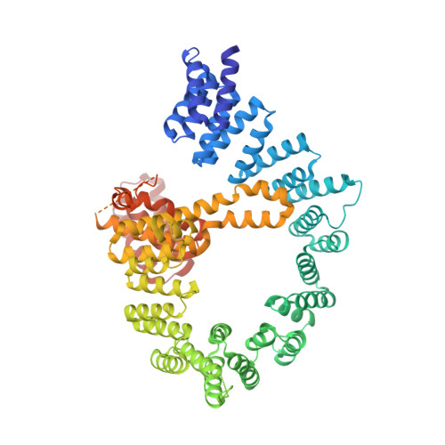
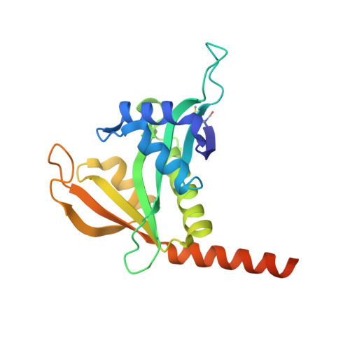
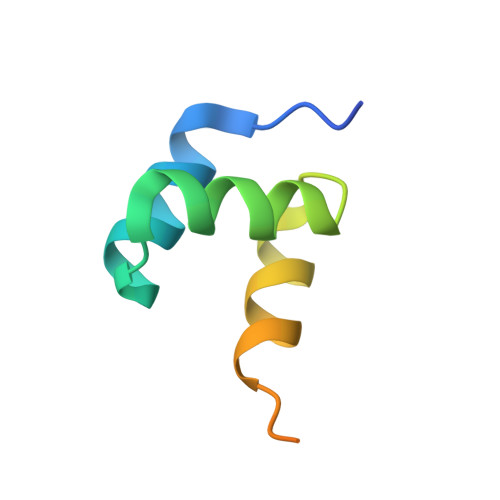
Structure of Naa15/Naa10 bound to HypK-THB


Experimental Data Snapshot

  • Method: X-RAY DIFFRACTION
  • Resolution: 2.60 Å
  • R-Value Free: 
    0.220 (Depositor), 0.220 (DCC) 
  • R-Value Work: 
    0.161 (Depositor), 0.170 (DCC) 
  • R-Value Observed: 
    0.164 (Depositor) 

wwPDB Validation   3D Report Full Report


Ligand Structure Quality Assessment 

Created with Raphaël 2.3.0Worse 01 BetterLigand structure goodness of fit to experimental dataBest fitted CMCClick on this verticalbar to view details

This is version 1.1 of the entry. See complete history


Literature

Structural basis of HypK regulating N-terminal acetylation by the NatA complex.

Weyer, F.A.Gumiero, A.Lapouge, K.Bange, G.Kopp, J.Sinning, I.

(2017) Nat Commun 8: 15726-15726

  • DOI: https://doi.org/10.1038/ncomms15726
  • Primary Citation of Related Structures:  
    5NNP, 5NNR

  • PubMed Abstract: 

    In eukaryotes, N-terminal acetylation is one of the most common protein modifications involved in a wide range of biological processes. Most N-acetyltransferase complexes (NATs) act co-translationally, with the heterodimeric NatA complex modifying the majority of substrate proteins. Here we show that the Huntingtin yeast two-hybrid protein K (HypK) binds tightly to the NatA complex comprising the auxiliary subunit Naa15 and the catalytic subunit Naa10. The crystal structures of NatA bound to HypK or to a N-terminal deletion variant of HypK were determined without or with a bi-substrate analogue, respectively. The HypK C-terminal region is responsible for high-affinity interaction with the C-terminal part of Naa15. In combination with acetylation assays, the HypK N-terminal region is identified as a negative regulator of the NatA acetylation activity. Our study provides mechanistic insights into the regulation of this pivotal protein modification.


  • Organizational Affiliation

    Heidelberg University Biochemistry Center (BZH), INF328, D-69120 Heidelberg, Germany.


Macromolecules
Find similar proteins by:  (by identity cutoff)  |  3D Structure
Entity ID: 1
MoleculeChains Sequence LengthOrganismDetailsImage
N-terminal acetyltransferase-like proteinA,
D [auth E]
745Thermochaetoides thermophila DSM 1495Mutation(s): 0 
Gene Names: CTHT_0031530
UniProt
Find proteins for G0S4M4 (Chaetomium thermophilum (strain DSM 1495 / CBS 144.50 / IMI 039719))
Explore G0S4M4 
Go to UniProtKB:  G0S4M4
Entity Groups  
Sequence Clusters30% Identity50% Identity70% Identity90% Identity95% Identity100% Identity
UniProt GroupG0S4M4
Sequence Annotations
Expand
  • Reference Sequence
Find similar proteins by:  (by identity cutoff)  |  3D Structure
Entity ID: 2
MoleculeChains Sequence LengthOrganismDetailsImage
Putative uncharacterized proteinB,
E [auth F]
195Thermochaetoides thermophila DSM 1495Mutation(s): 0 
Gene Names: CTHT_0063490
UniProt
Find proteins for G0SEE8 (Chaetomium thermophilum (strain DSM 1495 / CBS 144.50 / IMI 039719))
Explore G0SEE8 
Go to UniProtKB:  G0SEE8
Entity Groups  
Sequence Clusters30% Identity50% Identity70% Identity90% Identity95% Identity100% Identity
UniProt GroupG0SEE8
Sequence Annotations
Expand
  • Reference Sequence
Find similar proteins by:  (by identity cutoff)  |  3D Structure
Entity ID: 3
MoleculeChains Sequence LengthOrganismDetailsImage
Putative uncharacterized proteinC,
F [auth G]
62Thermochaetoides thermophila DSM 1495Mutation(s): 0 
Gene Names: CTHT_0058830
UniProt
Find proteins for G0SCY6 (Chaetomium thermophilum (strain DSM 1495 / CBS 144.50 / IMI 039719))
Explore G0SCY6 
Go to UniProtKB:  G0SCY6
Entity Groups  
Sequence Clusters30% Identity50% Identity70% Identity90% Identity95% Identity100% Identity
UniProt GroupG0SCY6
Sequence Annotations
Expand
  • Reference Sequence

Find similar proteins by:  Sequence   |   3D Structure  

Entity ID: 4
MoleculeChains Sequence LengthOrganismDetailsImage
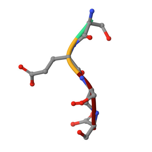
Ser-Glu-Ser-SerG [auth I],
H [auth L]
4synthetic constructMutation(s): 0 
Sequence Annotations
Expand
  • Reference Sequence
Small Molecules
Ligands 3 Unique
IDChains Name / Formula / InChI Key2D Diagram3D Interactions
CMC
Query on CMC

Download Ideal Coordinates CCD File 
NA [auth I],
OA [auth L]
CARBOXYMETHYL COENZYME *A
C23 H38 N7 O18 P3 S
OBUOSIHPWVNVJN-GRFIIANRSA-N
PO4
Query on PO4

Download Ideal Coordinates CCD File 
EA [auth E],
FA [auth E],
MA [auth F],
U [auth A]
PHOSPHATE ION
O4 P
NBIIXXVUZAFLBC-UHFFFAOYSA-K
GOL
Query on GOL

Download Ideal Coordinates CCD File 
AA [auth E]
BA [auth E]
CA [auth E]
DA [auth E]
GA [auth F]
GLYCEROL
C3 H8 O3
PEDCQBHIVMGVHV-UHFFFAOYSA-N
Modified Residues  1 Unique
IDChains TypeFormula2D DiagramParent
FME
Query on FME
B,
E [auth F]
L-PEPTIDE LINKINGC6 H11 N O3 SMET
Experimental Data & Validation

Experimental Data

  • Method: X-RAY DIFFRACTION
  • Resolution: 2.60 Å
  • R-Value Free:  0.220 (Depositor), 0.220 (DCC) 
  • R-Value Work:  0.161 (Depositor), 0.170 (DCC) 
  • R-Value Observed: 0.164 (Depositor) 
Space Group: P 1 21 1
Unit Cell:
Length ( Å )Angle ( ˚ )
a = 93.319α = 90
b = 106.041β = 94.98
c = 130.937γ = 90
Software Package:
Software NamePurpose
PHENIXrefinement
XDSdata reduction
Aimlessdata scaling
PHASERphasing

Structure Validation

View Full Validation Report


Ligand Structure Quality Assessment 

Created with Raphaël 2.3.0Worse 01 BetterLigand structure goodness of fit to experimental dataBest fitted CMCClick on this verticalbar to view details

Entry History & Funding Information

Deposition Data


Funding OrganizationLocationGrant Number
German Research FoundationGermanyFOR967
German Research FoundationGermanyGRK1188
German Research FoundationGermanyLeibniz Program

Revision History  (Full details and data files)

  • Version 1.0: 2017-06-14
    Type: Initial release
  • Version 1.1: 2024-11-13
    Changes: Author supporting evidence, Data collection, Database references, Structure summary