5NM7 | pdb_00005nm7

Crystal structure of Burkholderia AP3 phage endolysin


Experimental Data Snapshot

  • Method: X-RAY DIFFRACTION
  • Resolution: 1.72 Å
  • R-Value Free: 
    0.235 (Depositor), 0.266 (DCC) 
  • R-Value Work: 
    0.208 (Depositor) 
  • R-Value Observed: 
    0.210 (Depositor) 

wwPDB Validation 3D Report Full Report

Validation slider image for 5NM7

This is version 2.0 of the entry. See complete history

Literature

Modular endolysin of Burkholderia AP3 phage has the largest lysozyme-like catalytic subunit discovered to date and no catalytic aspartate residue.

Maciejewska, B.Zrubek, K.Espaillat, A.Wisniewska, M.Rembacz, K.P.Cava, F.Dubin, G.Drulis-Kawa, Z.

(2017) Sci Rep 7: 14501-14501

  • DOI: https://doi.org/10.1038/s41598-017-14797-9
  • Primary Citation Related Structures: 
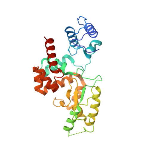
    5NM7

  • PubMed Abstract: 

    Endolysins are peptidoglycan-degrading enzymes utilized by bacteriophages to release the progeny from bacterial cells. The lytic properties of phage endolysins make them potential antibacterial agents for medical and industrial applications. Here, we present a comprehensive characterization of phage AP3 modular endolysin (AP3gp15) containing cell wall binding domain and an enzymatic domain (DUF3380 by BLASTP), both widespread and conservative. Our structural analysis demonstrates the low similarity of an enzymatic domain to known lysozymes and an unusual catalytic centre characterized by only a single glutamic acid residue and no aspartic acid. Thus, our findings suggest distinguishing a novel class of muralytic enzymes having the activity and catalytic centre organization of DUF3380. The lack of amino acid sequence homology between AP3gp15 and other known muralytic enzymes may reflect the evolutionary convergence of analogous glycosidases. Moreover, the broad antibacterial spectrum, lack of cytotoxic effect on human cells and the stability characteristics of AP3 endolysin advocate for its future application development.


  • Organizational Affiliation
    • Institute of Genetics and Microbiology, University of Wroclaw, Przybyszewskiego 63/77, 51-148, Wroclaw, Poland.

Macromolecule Content 

  • Total Structure Weight: 58.5 kDa 
  • Atom Count: 4,207 
  • Modeled Residue Count: 519 
  • Deposited Residue Count: 532 
  • Unique protein chains: 1

Macromolecules

Find similar proteins by:|  3D Structure
Entity ID: 1
MoleculeChains  Sequence LengthOrganismDetailsImage
Peptidoglycan-binding domain 1A [auth G],
B [auth A]
266BurkholderiaMutation(s): 0 
Gene Names: MYA_3986
UniProt
Find proteins for A0A1S5NV50 (Burkholderia phage AP3)
Explore A0A1S5NV50 
Go to UniProtKB:  A0A1S5NV50
Entity Groups
Sequence Clusters30% Identity50% Identity70% Identity90% Identity95% Identity100% Identity
UniProt GroupA0A1S5NV50
Sequence Annotations
Expand
Reference Sequence

Experimental Data & Validation

Experimental Data

  • Method: X-RAY DIFFRACTION
  • Resolution: 1.72 Å
  • R-Value Free:  0.235 (Depositor), 0.266 (DCC) 
  • R-Value Work:  0.208 (Depositor) 
  • R-Value Observed: 0.210 (Depositor) 
Space Group: H 3 2
Unit Cell:
Length ( Å )Angle ( ˚ )
a = 154.615α = 90
b = 154.615β = 90
c = 125.823γ = 120
Software Package:
Software NamePurpose
REFMACrefinement
iMOSFLMdata reduction
Aimlessdata scaling
MOLREPphasing

Structure Validation

View Full Validation Report


Entry History 

Deposition Data

Revision History  (Full details and data files)

  • Version 1.0: 2018-02-14
    Type: Initial release
  • Version 2.0: 2024-05-08
    Changes: Atomic model, Data collection, Database references, Refinement description