5NKW

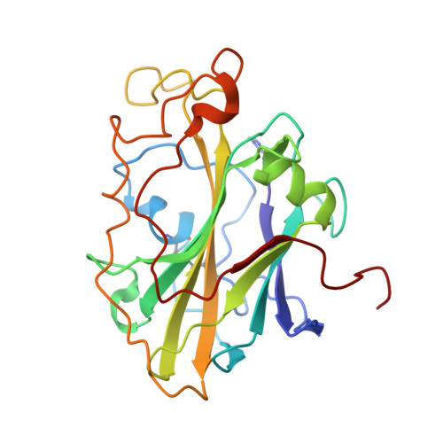
X-ray crystal structure of an AA9 LPMO


Experimental Data Snapshot

  • Method: X-RAY DIFFRACTION
  • Resolution: 1.48 Å
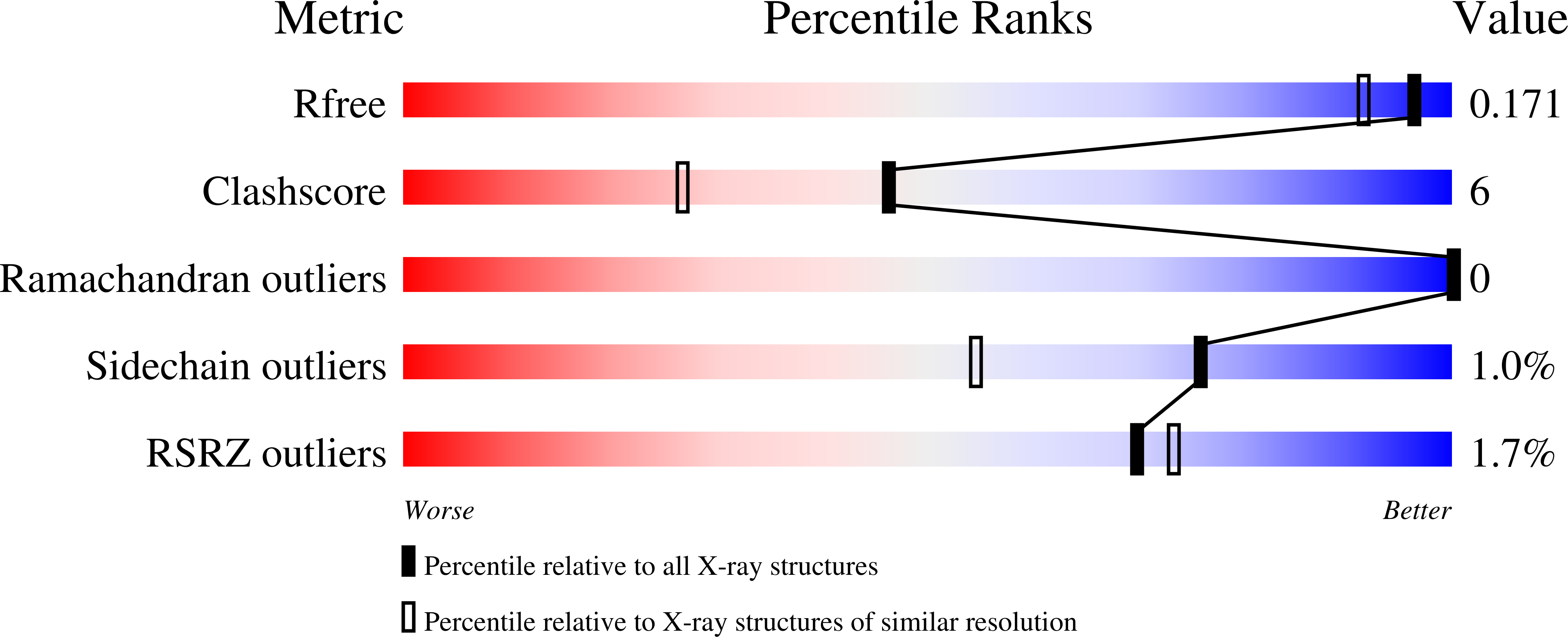
  • R-Value Free: 0.170 
  • R-Value Work: 0.123 
  • R-Value Observed: 0.126 

Starting Model: experimental
View more details

wwPDB Validation   3D Report Full Report


Ligand Structure Quality Assessment 


This is version 2.1 of the entry. See complete history


Literature

Structural and electronic determinants of lytic polysaccharide monooxygenase reactivity on polysaccharide substrates.

Simmons, T.J.Frandsen, K.E.H.Ciano, L.Tryfona, T.Lenfant, N.Poulsen, J.C.Wilson, L.F.L.Tandrup, T.Tovborg, M.Schnorr, K.Johansen, K.S.Henrissat, B.Walton, P.H.Lo Leggio, L.Dupree, P.

(2017) Nat Commun 8: 1064-1064

  • DOI: https://doi.org/10.1038/s41467-017-01247-3
  • Primary Citation of Related Structures:  
    5NKW, 5NLN, 5NLO, 5NLP, 5NLQ, 5NLR, 5NLS, 5NLT

  • PubMed Abstract: 

    Lytic polysaccharide monooxygenases (LPMOs) are industrially important copper-dependent enzymes that oxidatively cleave polysaccharides. Here we present a functional and structural characterization of two closely related AA9-family LPMOs from Lentinus similis (LsAA9A) and Collariella virescens (CvAA9A). LsAA9A and CvAA9A cleave a range of polysaccharides, including cellulose, xyloglucan, mixed-linkage glucan and glucomannan. LsAA9A additionally cleaves isolated xylan substrates. The structures of CvAA9A and of LsAA9A bound to cellulosic and non-cellulosic oligosaccharides provide insight into the molecular determinants of their specificity. Spectroscopic measurements reveal differences in copper co-ordination upon the binding of xylan and glucans. LsAA9A activity is less sensitive to the reducing agent potential when cleaving xylan, suggesting that distinct catalytic mechanisms exist for xylan and glucan cleavage. Overall, these data show that AA9 LPMOs can display different apparent substrate specificities dependent upon both productive protein-carbohydrate interactions across a binding surface and also electronic considerations at the copper active site.


  • Organizational Affiliation

    Department of Biochemistry, University of Cambridge, Cambridge, CB2 1QW, UK.


Macromolecules
Find similar proteins by:  (by identity cutoff)  |  3D Structure
Entity ID: 1
MoleculeChains Sequence LengthOrganismDetailsImage
Auxiliary activity 9235Panus similisMutation(s): 0 
EC: 1.14.99.56
UniProt
Find proteins for A0A0S2GKZ1 (Panus similis)
Explore A0A0S2GKZ1 
Go to UniProtKB:  A0A0S2GKZ1
Entity Groups  
Sequence Clusters30% Identity50% Identity70% Identity90% Identity95% Identity100% Identity
UniProt GroupA0A0S2GKZ1
Glycosylation
Glycosylation Sites: 1
Sequence Annotations
Expand
  • Reference Sequence
Oligosaccharides

Help

Entity ID: 2
MoleculeChains Length2D Diagram Glycosylation3D Interactions
beta-D-glucopyranose-(1-4)-beta-D-mannopyranose-(1-4)-beta-D-glucopyranose-(1-4)-beta-D-glucopyranose-(1-4)-beta-D-mannopyranose-(1-4)-beta-D-glucopyranose-(1-4)-beta-D-mannopyranose
B
7N/A
Glycosylation Resources
GlyTouCan:  G60546OC
GlyCosmos:  G60546OC
Entity ID: 3
MoleculeChains Length2D Diagram Glycosylation3D Interactions
beta-D-glucopyranose-(1-4)-beta-D-mannopyranose-(1-4)-beta-D-mannopyranose
C
3N/A
Glycosylation Resources
GlyTouCan:  G91016IX
GlyCosmos:  G91016IX
GlyGen:  G91016IX
Small Molecules
Ligands 5 Unique
IDChains Name / Formula / InChI Key2D Diagram3D Interactions
NAG
Query on NAG

Download Ideal Coordinates CCD File 
E [auth A]2-acetamido-2-deoxy-beta-D-glucopyranose
C8 H15 N O6
OVRNDRQMDRJTHS-FMDGEEDCSA-N
BMA
Query on BMA

Download Ideal Coordinates CCD File 
X [auth A]beta-D-mannopyranose
C6 H12 O6
WQZGKKKJIJFFOK-RWOPYEJCSA-N
BGC
Query on BGC

Download Ideal Coordinates CCD File 
W [auth A]beta-D-glucopyranose
C6 H12 O6
WQZGKKKJIJFFOK-VFUOTHLCSA-N
CU
Query on CU

Download Ideal Coordinates CCD File 
D [auth A]COPPER (II) ION
Cu
JPVYNHNXODAKFH-UHFFFAOYSA-N
CL
Query on CL

Download Ideal Coordinates CCD File 
F [auth A]
G [auth A]
H [auth A]
I [auth A]
J [auth A]
F [auth A],
G [auth A],
H [auth A],
I [auth A],
J [auth A],
K [auth A],
L [auth A],
M [auth A],
N [auth A],
O [auth A],
P [auth A],
Q [auth A],
R [auth A],
S [auth A],
T [auth A],
U [auth A],
V [auth A]
CHLORIDE ION
Cl
VEXZGXHMUGYJMC-UHFFFAOYSA-M
Modified Residues  1 Unique
IDChains TypeFormula2D DiagramParent
HIC
Query on HIC
A
L-PEPTIDE LINKINGC7 H11 N3 O2HIS
Experimental Data & Validation

Experimental Data

  • Method: X-RAY DIFFRACTION
  • Resolution: 1.48 Å
  • R-Value Free: 0.170 
  • R-Value Work: 0.123 
  • R-Value Observed: 0.126 
  • Space Group: P 41 3 2
Unit Cell:
Length ( Å )Angle ( ˚ )
a = 125.29α = 90
b = 125.29β = 90
c = 125.29γ = 90
Software Package:
Software NamePurpose
REFMACrefinement
XDSdata reduction
XSCALEdata scaling
REFMACphasing

Structure Validation

View Full Validation Report


Ligand Structure Quality Assessment 


Entry History & Funding Information

Deposition Data


Funding OrganizationLocationGrant Number
Danish Council for Strategic ResearchDenmark12-134923

Revision History  (Full details and data files)

  • Version 1.0: 2017-11-01
    Type: Initial release
  • Version 2.0: 2020-07-29
    Type: Remediation
    Reason: Carbohydrate remediation
    Changes: Atomic model, Data collection, Derived calculations, Structure summary
  • Version 2.1: 2024-01-17
    Changes: Data collection, Database references, Derived calculations, Refinement description, Structure summary