5N07 | pdb_00005n07

Structure of the [4Fe-4S] form of the NO response regulator NsrR


Experimental Data Snapshot

  • Method: X-RAY DIFFRACTION
  • Resolution: 1.95 Å
  • R-Value Free: 
    0.227 (Depositor), 0.230 (DCC) 
  • R-Value Work: 
    0.195 (Depositor), 0.200 (DCC) 
  • R-Value Observed: 
    0.196 (Depositor) 

wwPDB Validation 3D Report Full Report

Validation slider image for 5N07

This is version 1.2 of the entry. See complete history

Literature

Crystal structures of the NO sensor NsrR reveal how its iron-sulfur cluster modulates DNA binding.

Volbeda, A.Dodd, E.L.Darnault, C.Crack, J.C.Renoux, O.Hutchings, M.I.Le Brun, N.E.Fontecilla-Camps, J.C.

(2017) Nat Commun 8: 15052-15052

  • DOI: https://doi.org/10.1038/ncomms15052
  • Primary Citation Related Structures: 
    5N07, 5N08

  • PubMed Abstract: 

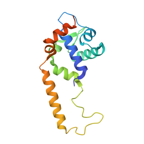
    NsrR from Streptomyces coelicolor (Sc) regulates the expression of three genes through the progressive degradation of its [4Fe-4S] cluster on nitric oxide (NO) exposure. We report the 1.95 Å resolution crystal structure of dimeric holo-ScNsrR and show that the cluster is coordinated by the three invariant Cys residues from one monomer and, unexpectedly, Asp8 from the other. A cavity map suggests that NO displaces Asp8 as a cluster ligand and, while D8A and D8C variants remain NO sensitive, DNA binding is affected. A structural comparison of holo-ScNsrR with an apo-IscR-DNA complex shows that the [4Fe-4S] cluster stabilizes a turn between ScNsrR Cys93 and Cys99 properly oriented to interact with the DNA backbone. In addition, an apo ScNsrR structure suggests that Asn97 from this turn, along with Arg12, which forms a salt-bridge with Asp8, are instrumental in modulating the position of the DNA recognition helix region relative to its major groove.


  • Organizational Affiliation
    • Metalloproteins Unit, Institut de Biologie Structurale, CEA, CNRS, Université Grenoble-Alpes, 71, avenue des Martyrs, CS 10090, 38044 Grenoble cedex 9, France.

Macromolecule Content 

  • Total Structure Weight: 17.99 kDa 
  • Atom Count: 1,061 
  • Modeled Residue Count: 137 
  • Deposited Residue Count: 161 
  • Unique protein chains: 1

Macromolecules

Find similar proteins by:|  3D Structure
Entity ID: 1
MoleculeChains  Sequence LengthOrganismDetailsImage
HTH-type transcriptional repressor NsrR161Streptomyces coelicolor A3(2)Mutation(s): 0 
Gene Names: nsrRSCO7427SC6D11.23
UniProt
Find proteins for Q9L132 (Streptomyces coelicolor (strain ATCC BAA-471 / A3(2) / M145))
Explore Q9L132 
Go to UniProtKB:  Q9L132
Entity Groups
Sequence Clusters30% Identity50% Identity70% Identity90% Identity95% Identity100% Identity
UniProt GroupQ9L132
Sequence Annotations
Expand
Reference Sequence

Experimental Data & Validation

Experimental Data

  • Method: X-RAY DIFFRACTION
  • Resolution: 1.95 Å
  • R-Value Free:  0.227 (Depositor), 0.230 (DCC) 
  • R-Value Work:  0.195 (Depositor), 0.200 (DCC) 
  • R-Value Observed: 0.196 (Depositor) 
Space Group: P 65 2 2
Unit Cell:
Length ( Å )Angle ( ˚ )
a = 78.76α = 90
b = 78.76β = 90
c = 93.26γ = 120
Software Package:
Software NamePurpose
REFMACrefinement
XDSdata reduction
Aimlessdata scaling
PHENIXphasing

Structure Validation

View Full Validation Report


Entry History 

Deposition Data

Revision History  (Full details and data files)

  • Version 1.0: 2017-04-26
    Type: Initial release
  • Version 1.1: 2017-05-03
    Changes: Database references
  • Version 1.2: 2024-05-08
    Changes: Data collection, Database references, Derived calculations