5LTR | pdb_00005ltr

Structure of the Yellow-Green Fluorescent Protein mNeonGreen from Branchiostoma lanceolatum at the near physiological pH 8.0


Experimental Data Snapshot

  • Method: X-RAY DIFFRACTION
  • Resolution: 1.21 Å
  • R-Value Free: 
    0.184 (Depositor), 0.191 (DCC) 
  • R-Value Work: 
    0.158 (Depositor), 0.167 (DCC) 
  • R-Value Observed: 
    0.159 (Depositor) 

Starting Model: experimental
View more details

wwPDB Validation 3D Report Full Report

Validation slider image for 5LTR

This is version 1.2 of the entry. See complete history

Literature

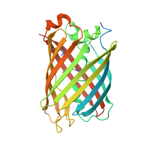
Structural analysis of the bright monomeric yellow-green fluorescent protein mNeonGreen obtained by directed evolution.

Clavel, D.Gotthard, G.von Stetten, D.De Sanctis, D.Pasquier, H.Lambert, G.G.Shaner, N.C.Royant, A.

(2016) Acta Crystallogr D Struct Biol 72: 1298-1307

  • DOI: https://doi.org/10.1107/S2059798316018623
  • Primary Citation Related Structures: 
    5LTP, 5LTQ, 5LTR

  • PubMed Abstract: 

    Until recently, genes coding for homologues of the autofluorescent protein GFP had only been identified in marine organisms from the phyla Cnidaria and Arthropoda. New fluorescent-protein genes have now been found in the phylum Chordata, coding for particularly bright oligomeric fluorescent proteins such as the tetrameric yellow fluorescent protein lanYFP from Branchiostoma lanceolatum. A successful monomerization attempt led to the development of the bright yellow-green fluorescent protein mNeonGreen. The structures of lanYFP and mNeonGreen have been determined and compared in order to rationalize the directed evolution process leading from a bright, tetrameric to a still bright, monomeric fluorescent protein. An unusual discolouration of crystals of mNeonGreen was observed after X-ray data collection, which was investigated using a combination of X-ray crystallography and UV-visible absorption and Raman spectroscopies, revealing the effects of specific radiation damage in the chromophore cavity. It is shown that X-rays rapidly lead to the protonation of the phenolate O atom of the chromophore and to the loss of its planarity at the methylene bridge.


  • Organizational Affiliation
    • Université Grenoble Alpes, Institut de Biologie Structurale (IBS), F-38044 Grenoble, France.

Macromolecule Content 

  • Total Structure Weight: 30.73 kDa 
  • Atom Count: 2,107 
  • Modeled Residue Count: 217 
  • Deposited Residue Count: 269 
  • Unique protein chains: 1

Macromolecules

Find similar proteins by:|  3D Structure
Entity ID: 1
MoleculeChains  Sequence LengthOrganismDetailsImage
mNeonGreen269Branchiostoma lanceolatumMutation(s): 0 
Gene Names: blFP-Y3
UniProt
Find proteins for B1PNC0 (Branchiostoma lanceolatum)
Explore B1PNC0 
Go to UniProtKB:  B1PNC0
Entity Groups
Sequence Clusters30% Identity50% Identity70% Identity90% Identity95% Identity100% Identity
UniProt GroupB1PNC0
Sequence Annotations
Expand
Reference Sequence

Small Molecules

Ligands 1 Unique
IDChains Name / Formula / InChI Key2D Diagram3D Interactions
CL

Query on CL


Download:Ideal Coordinates CCD File
B [auth A]CHLORIDE ION
Cl
VEXZGXHMUGYJMC-UHFFFAOYSA-M
Modified Residues  2 Unique
IDChains TypeFormula2D DiagramParent
CR2
Query on CR2
A
L-PEPTIDE LINKINGC13 H13 N3 O4GLY, TYR, GLY
KCX
Query on KCX
A
L-PEPTIDE LINKINGC7 H14 N2 O4LYS

Experimental Data & Validation

Experimental Data

  • Method: X-RAY DIFFRACTION
  • Resolution: 1.21 Å
  • R-Value Free:  0.184 (Depositor), 0.191 (DCC) 
  • R-Value Work:  0.158 (Depositor), 0.167 (DCC) 
  • R-Value Observed: 0.159 (Depositor) 
Space Group: P 65 2 2
Unit Cell:
Length ( Å )Angle ( ˚ )
a = 72.27α = 90
b = 72.27β = 90
c = 153.97γ = 120
Software Package:
Software NamePurpose
XDSdata reduction
XSCALEdata scaling
PHASERphasing
REFMACrefinement

Structure Validation

View Full Validation Report


Entry History 

Deposition Data

Revision History  (Full details and data files)

  • Version 1.0: 2016-12-07
    Type: Initial release
  • Version 1.1: 2016-12-21
    Changes: Database references
  • Version 1.2: 2024-01-17
    Changes: Data collection, Database references, Refinement description