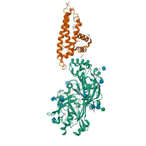
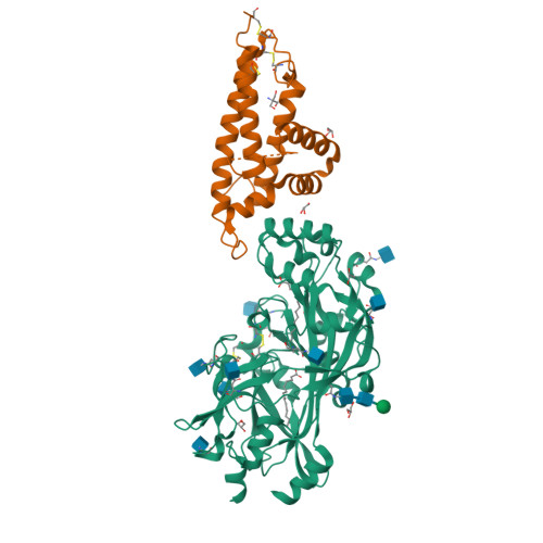
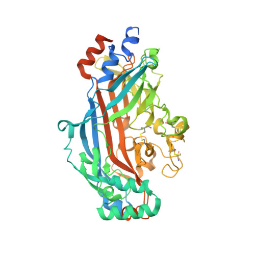
5LGD

The CIDRa domain from MCvar1 PfEMP1 bound to CD36


Experimental Data Snapshot

  • Method: X-RAY DIFFRACTION
  • Resolution: 2.07 Å
  • R-Value Free: 0.253 
  • R-Value Work: 0.212 
  • R-Value Observed: 0.214 

Starting Model: experimental
View more details

wwPDB Validation   3D Report Full Report


Ligand Structure Quality Assessment 


This is version 2.1 of the entry. See complete history


Literature

The structural basis for CD36 binding by the malaria parasite.

Hsieh, F.L.Turner, L.Bolla, J.R.Robinson, C.V.Lavstsen, T.Higgins, M.K.

(2016) Nat Commun 7: 12837-12837

  • DOI: https://doi.org/10.1038/ncomms12837
  • Primary Citation of Related Structures:  
    5LGD

  • PubMed Abstract: 

    CD36 is a scavenger receptor involved in fatty acid metabolism, innate immunity and angiogenesis. It interacts with lipoprotein particles and facilitates uptake of long chain fatty acids. It is also the most common target of the PfEMP1 proteins of the malaria parasite, Plasmodium falciparum, tethering parasite-infected erythrocytes to endothelial receptors. This prevents their destruction by splenic clearance and allows increased parasitaemia. Here we describe the structure of CD36 in complex with long chain fatty acids and a CD36-binding PfEMP1 protein domain. A conserved hydrophobic pocket allows the hugely diverse PfEMP1 protein family to bind to a conserved phenylalanine residue at the membrane distal tip of CD36. This phenylalanine is also required for CD36 to interact with lipoprotein particles. By targeting a site on CD36 that is required for its physiological function, PfEMP1 proteins maintain the ability to tether to the endothelium and avoid splenic clearance.


  • Organizational Affiliation

    Department of Biochemistry, University of Oxford, South Parks Road, Oxford OX1 3QU, UK.


Macromolecules
Find similar proteins by:  (by identity cutoff)  |  3D Structure
Entity ID: 1
MoleculeChains Sequence LengthOrganismDetailsImage
Platelet glycoprotein 4472Homo sapiensMutation(s): 0 
Gene Names: CD36GP3BGP4
UniProt & NIH Common Fund Data Resources
Find proteins for P16671 (Homo sapiens)
Explore P16671 
Go to UniProtKB:  P16671
PHAROS:  P16671
GTEx:  ENSG00000135218 
Entity Groups  
Sequence Clusters30% Identity50% Identity70% Identity90% Identity95% Identity100% Identity
UniProt GroupP16671
Glycosylation
Glycosylation Sites: 9Go to GlyGen: P16671-1
Sequence Annotations
Expand
  • Reference Sequence
Find similar proteins by:  (by identity cutoff)  |  3D Structure
Entity ID: 2
MoleculeChains Sequence LengthOrganismDetailsImage
PfEMP1 variant 1 of strain MC179Plasmodium falciparumMutation(s): 0 
Gene Names: MCvar-1 PfEMP1
UniProt
Find proteins for Q25733 (Plasmodium falciparum)
Explore Q25733 
Go to UniProtKB:  Q25733
Entity Groups  
Sequence Clusters30% Identity50% Identity70% Identity90% Identity95% Identity100% Identity
UniProt GroupQ25733
Sequence Annotations
Expand
  • Reference Sequence
Oligosaccharides

Help

Entity ID: 3
MoleculeChains Length2D Diagram Glycosylation3D Interactions
alpha-D-mannopyranose-(1-4)-2-acetamido-2-deoxy-beta-D-glucopyranose-(1-4)-2-acetamido-2-deoxy-beta-D-glucopyranose
C
3N-Glycosylation
Glycosylation Resources
GlyTouCan:  G62182OO
GlyCosmos:  G62182OO
GlyGen:  G62182OO
Small Molecules
Ligands 4 Unique
IDChains Name / Formula / InChI Key2D Diagram3D Interactions
PLM
Query on PLM

Download Ideal Coordinates CCD File 
L [auth A],
M [auth A]
PALMITIC ACID
C16 H32 O2
IPCSVZSSVZVIGE-UHFFFAOYSA-N
NAG
Query on NAG

Download Ideal Coordinates CCD File 
D [auth A]
E [auth A]
F [auth A]
G [auth A]
H [auth A]
D [auth A],
E [auth A],
F [auth A],
G [auth A],
H [auth A],
I [auth A],
J [auth A],
K [auth A]
2-acetamido-2-deoxy-beta-D-glucopyranose
C8 H15 N O6
OVRNDRQMDRJTHS-FMDGEEDCSA-N
TRS
Query on TRS

Download Ideal Coordinates CCD File 
R [auth A],
T [auth B]
2-AMINO-2-HYDROXYMETHYL-PROPANE-1,3-DIOL
C4 H12 N O3
LENZDBCJOHFCAS-UHFFFAOYSA-O
GOL
Query on GOL

Download Ideal Coordinates CCD File 
N [auth A],
O [auth A],
P [auth A],
Q [auth A],
S [auth B]
GLYCEROL
C3 H8 O3
PEDCQBHIVMGVHV-UHFFFAOYSA-N
Experimental Data & Validation

Experimental Data

  • Method: X-RAY DIFFRACTION
  • Resolution: 2.07 Å
  • R-Value Free: 0.253 
  • R-Value Work: 0.212 
  • R-Value Observed: 0.214 
  • Space Group: C 1 2 1
Unit Cell:
Length ( Å )Angle ( ˚ )
a = 129.826α = 90
b = 40.726β = 114.85
c = 138.836γ = 90
Software Package:
Software NamePurpose
BUSTERrefinement
XDSdata reduction
XDSdata scaling
PHASERphasing

Structure Validation

View Full Validation Report


Ligand Structure Quality Assessment 


Entry History & Funding Information

Deposition Data


Funding OrganizationLocationGrant Number
Wellcome TrustUnited Kingdom101020/Z/13/Z

Revision History  (Full details and data files)

  • Version 1.0: 2016-08-31
    Type: Initial release
  • Version 1.1: 2016-10-05
    Changes: Database references
  • Version 1.2: 2019-10-16
    Changes: Data collection
  • Version 2.0: 2020-07-29
    Type: Remediation
    Reason: Carbohydrate remediation
    Changes: Atomic model, Data collection, Derived calculations, Structure summary
  • Version 2.1: 2024-01-10
    Changes: Data collection, Database references, Refinement description, Structure summary