5L43 | pdb_00005l43

Structure of K26-DCP


Experimental Data Snapshot

  • Method: X-RAY DIFFRACTION
  • Resolution: 1.80 Å
  • R-Value Free: 
    0.200 (Depositor), 0.209 (DCC) 
  • R-Value Work: 
    0.177 (Depositor), 0.186 (DCC) 
  • R-Value Observed: 
    0.178 (Depositor) 

Starting Model: experimental
View more details

wwPDB Validation 3D Report Full Report

Validation slider image for 5L43

This is version 1.4 of the entry. See complete history

Literature

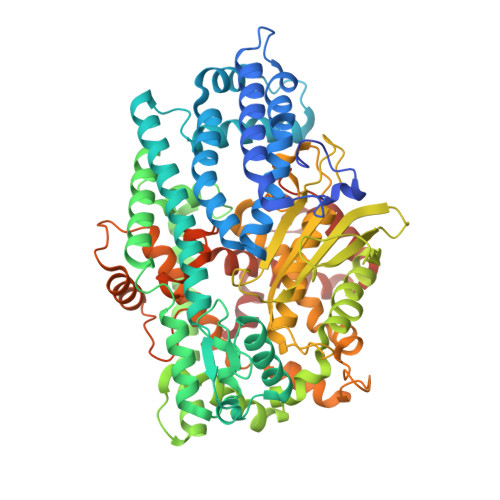
Crystal structure of a peptidyl-dipeptidase K-26-DCP from Actinomycete in complex with its natural inhibitor.

Masuyer, G.Cozier, G.E.Kramer, G.J.Bachmann, B.O.Acharya, K.R.

(2016) FEBS J 283: 4357-4369

  • DOI: https://doi.org/10.1111/febs.13928
  • Primary Citation Related Structures: 
    5L43, 5L44

  • PubMed Abstract: 

    Several soil-derived Actinobacteria produce secondary metabolites that are proven specific and potent inhibitors of the human angiotensin-I-converting enzyme (ACE), a key target for the modulation of hypertension through its role in the renin-angiotensin-aldosterone system. K-26-DCP is a zinc dipeptidyl carboxypeptidase (DCP) produced by Astrosporangium hypotensionis, and an ancestral homologue of ACE. Here we report the high-resolution crystal structures of K-26-DCP and of its complex with the natural microbial tripeptide product K-26. The experimental results provide the structural basis for better understanding the specificity of K-26 for human ACE over bacterial DCPs. Structural data are available in the PDB under the accession numbers 5L43 and 5L44.


  • Organizational Affiliation
    • Department of Biology and Biochemistry, University of Bath, UK.

Macromolecule Content 

  • Total Structure Weight: 153.76 kDa 
  • Atom Count: 11,418 
  • Modeled Residue Count: 1,324 
  • Deposited Residue Count: 1,366 
  • Unique protein chains: 1

Macromolecules

Find similar proteins by:|  3D Structure
Entity ID: 1
MoleculeChains  Sequence LengthOrganismDetailsImage
K-26 dipeptidyl carboxypeptidase
A, B
683Astrosporangium hypotensionis K-26Mutation(s): 0 
EC: 3.4.15.5
UniProt
Find proteins for A0A1L1QK30 (Astrosporangium hypotensionis K-26)
Explore A0A1L1QK30 
Go to UniProtKB:  A0A1L1QK30
Entity Groups
Sequence Clusters30% Identity50% Identity70% Identity90% Identity95% Identity100% Identity
UniProt GroupA0A1L1QK30
Sequence Annotations
Expand
Reference Sequence

Small Molecules

Ligands 3 Unique
IDChains Name / Formula / InChI Key2D Diagram3D Interactions
SO4

Query on SO4


Download:Ideal Coordinates CCD File
E [auth A],
F [auth A],
I [auth B],
J [auth B]
SULFATE ION
O4 S
QAOWNCQODCNURD-UHFFFAOYSA-L
ZN

Query on ZN


Download:Ideal Coordinates CCD File
C [auth A],
G [auth B]
ZINC ION
Zn
PTFCDOFLOPIGGS-UHFFFAOYSA-N
MG

Query on MG


Download:Ideal Coordinates CCD File
D [auth A],
H [auth B]
MAGNESIUM ION
Mg
JLVVSXFLKOJNIY-UHFFFAOYSA-N

Experimental Data & Validation

Experimental Data

  • Method: X-RAY DIFFRACTION
  • Resolution: 1.80 Å
  • R-Value Free:  0.200 (Depositor), 0.209 (DCC) 
  • R-Value Work:  0.177 (Depositor), 0.186 (DCC) 
  • R-Value Observed: 0.178 (Depositor) 
Space Group: P 1
Unit Cell:
Length ( Å )Angle ( ˚ )
a = 61.47α = 97.34
b = 66.936β = 90.86
c = 101.786γ = 117.31
Software Package:
Software NamePurpose
REFMACrefinement
DIALSdata reduction
Aimlessdata scaling
PHASERphasing

Structure Validation

View Full Validation Report


Entry History 

& Funding Information

Deposition Data


Funding OrganizationLocationGrant Number
Medical Research Council (United Kingdom)United KingdomMR/M026647/1

Revision History  (Full details and data files)

  • Version 1.0: 2016-10-26
    Type: Initial release
  • Version 1.1: 2016-11-02
    Changes: Database references
  • Version 1.2: 2016-12-14
    Changes: Database references
  • Version 1.3: 2017-09-13
    Changes: Author supporting evidence
  • Version 1.4: 2024-01-10
    Changes: Data collection, Database references, Derived calculations, Refinement description