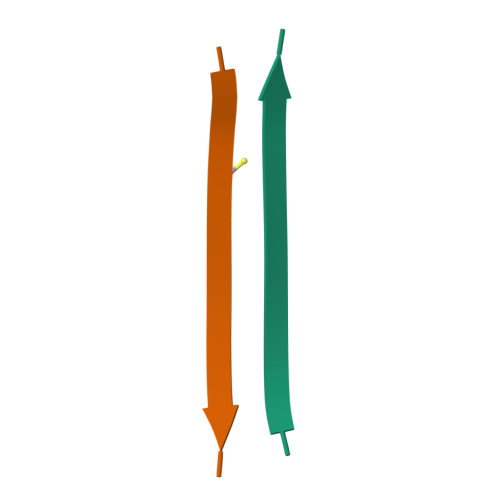
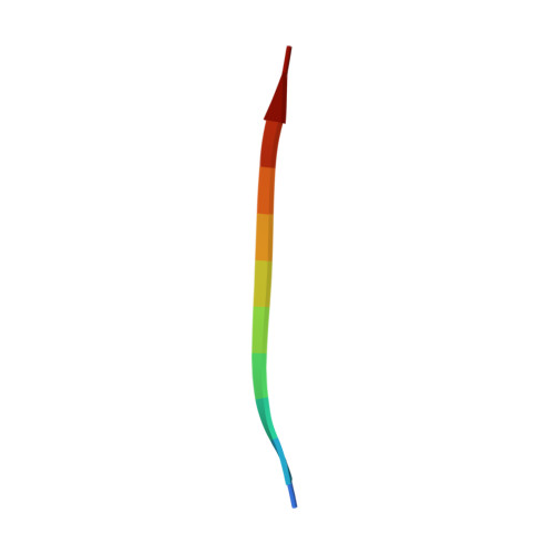
Atomic structures of fibrillar segments of hIAPP suggest tightly mated beta-sheets are important for cytotoxicity.
Krotee, P., Rodriguez, J.A., Sawaya, M.R., Cascio, D., Reyes, F.E., Shi, D., Hattne, J., Nannenga, B.L., Oskarsson, M.E., Philipp, S., Griner, S., Jiang, L., Glabe, C.G., Westermark, G.T., Gonen, T., Eisenberg, D.S.(2017) Elife 6
- PubMed: 28045370
- DOI: https://doi.org/10.7554/eLife.19273
- Primary Citation of Related Structures:
5KNZ, 5KO0 - PubMed Abstract:
hIAPP fibrils are associated with Type-II Diabetes, but the link of hIAPP structure to islet cell death remains elusive. Here we observe that hIAPP fibrils are cytotoxic to cultured pancreatic β-cells, leading us to determine the structure and cytotoxicity of protein segments composing the amyloid spine of hIAPP. Using the cryoEM method MicroED, we discover that one segment, 19-29 S20G, forms pairs of β-sheets mated by a dry interface that share structural features with and are similarly cytotoxic to full-length hIAPP fibrils. In contrast, a second segment, 15-25 WT, forms non-toxic labile β-sheets. These segments possess different structures and cytotoxic effects, however, both can seed full-length hIAPP, and cause hIAPP to take on the cytotoxic and structural features of that segment. These results suggest that protein segment structures represent polymorphs of their parent protein and that segment 19-29 S20G may serve as a model for the toxic spine of hIAPP.
Organizational Affiliation:
Department of Biological Chemistry, Howard Hughes Medical Institute, University of California, Los Angeles, Los Angeles, United States.


