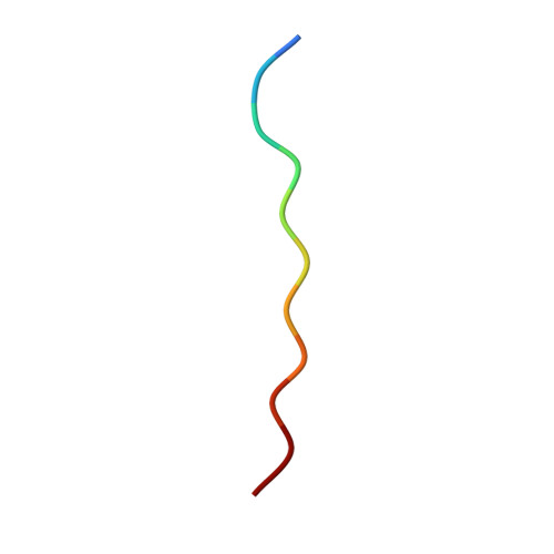
5KNZ

Human Islet Amyloid Polypeptide Segment 19-SGNNFGAILSS-29 with Early Onset S20G Mutation Determined by MicroED


Experimental Data Snapshot

  • Method: ELECTRON CRYSTALLOGRAPHY
  • Resolution: 1.90 Å
  • R-Value Free: 0.275 
  • R-Value Work: 0.228 
  • R-Value Observed: 0.232 

wwPDB Validation   3D Report Full Report


This is version 1.7 of the entry. See complete history


Literature

Atomic structures of fibrillar segments of hIAPP suggest tightly mated beta-sheets are important for cytotoxicity.

Krotee, P.Rodriguez, J.A.Sawaya, M.R.Cascio, D.Reyes, F.E.Shi, D.Hattne, J.Nannenga, B.L.Oskarsson, M.E.Philipp, S.Griner, S.Jiang, L.Glabe, C.G.Westermark, G.T.Gonen, T.Eisenberg, D.S.

(2017) Elife 6

  • DOI: https://doi.org/10.7554/eLife.19273
  • Primary Citation of Related Structures:  
    5KNZ, 5KO0

  • PubMed Abstract: 

    hIAPP fibrils are associated with Type-II Diabetes, but the link of hIAPP structure to islet cell death remains elusive. Here we observe that hIAPP fibrils are cytotoxic to cultured pancreatic β-cells, leading us to determine the structure and cytotoxicity of protein segments composing the amyloid spine of hIAPP. Using the cryoEM method MicroED, we discover that one segment, 19-29 S20G, forms pairs of β-sheets mated by a dry interface that share structural features with and are similarly cytotoxic to full-length hIAPP fibrils. In contrast, a second segment, 15-25 WT, forms non-toxic labile β-sheets. These segments possess different structures and cytotoxic effects, however, both can seed full-length hIAPP, and cause hIAPP to take on the cytotoxic and structural features of that segment. These results suggest that protein segment structures represent polymorphs of their parent protein and that segment 19-29 S20G may serve as a model for the toxic spine of hIAPP.


  • Organizational Affiliation

    Department of Biological Chemistry, Howard Hughes Medical Institute, University of California, Los Angeles, Los Angeles, United States.


Macromolecules

Find similar proteins by:  Sequence   |   3D Structure  

Entity ID: 1
MoleculeChains Sequence LengthOrganismDetailsImage
hIAPP(residues 19-29)S20G11Homo sapiensMutation(s): 1 
UniProt & NIH Common Fund Data Resources
Find proteins for P10997 (Homo sapiens)
Explore P10997 
Go to UniProtKB:  P10997
PHAROS:  P10997
GTEx:  ENSG00000121351 
Entity Groups  
Sequence Clusters30% Identity50% Identity70% Identity90% Identity95% Identity100% Identity
UniProt GroupP10997
Sequence Annotations
Expand
  • Reference Sequence
Experimental Data & Validation

Experimental Data

  • Method: ELECTRON CRYSTALLOGRAPHY
  • Resolution: 1.90 Å
  • R-Value Free: 0.275 
  • R-Value Work: 0.228 
  • R-Value Observed: 0.232 
  • Space Group: P 21 21 21
Unit Cell:
Length ( Å )Angle ( ˚ )
a = 4.78α = 90
b = 18.6β = 90
c = 70.8γ = 90
Software Package:
Software NamePurpose
XSCALEdata scaling
PHASERphasing
PHENIXrefinement
PDB_EXTRACTdata extraction
EM Software:
TaskSoftware PackageVersion
MODEL REFINEMENTPHENIX1.9

Structure Validation

View Full Validation Report


Entry History & Funding Information

Deposition Data


Funding OrganizationLocationGrant Number
National Institutes of Health/National Institute on Aging (NIH/NIA)United StatesR01 AG029430
Howard Hughes Medical Institute (HHMI)United States--

Revision History  (Full details and data files)

  • Version 1.0: 2016-12-21
    Type: Initial release
  • Version 1.1: 2017-09-13
    Changes: Author supporting evidence, Data collection
  • Version 1.2: 2018-04-25
    Changes: Data collection
  • Version 1.3: 2018-06-06
    Changes: Data collection, Experimental preparation, Refinement description
  • Version 1.4: 2019-09-04
    Changes: Data collection, Database references
  • Version 1.5: 2019-11-20
    Changes: Author supporting evidence
  • Version 1.6: 2021-06-30
    Changes: Data collection
  • Version 1.7: 2024-03-06
    Changes: Data collection, Database references, Refinement description