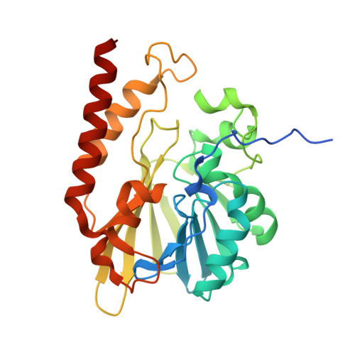
5K0W

Crystal structure of the metallo-beta-lactamase GOB-18 from Elizabethkingia meningoseptica


Experimental Data Snapshot

  • Method: X-RAY DIFFRACTION
  • Resolution: 2.61 Å
  • R-Value Free: 0.242 
  • R-Value Work: 0.192 
  • R-Value Observed: 0.195 

Starting Model: experimental
View more details

wwPDB Validation   3D Report Full Report


This is version 1.4 of the entry. See complete history


Literature

Crystal Structure of the Metallo-beta-Lactamase GOB in the Periplasmic Dizinc Form Reveals an Unusual Metal Site.

Moran-Barrio, J.Lisa, M.N.Larrieux, N.Drusin, S.I.Viale, A.M.Moreno, D.M.Buschiazzo, A.Vila, A.J.

(2016) Antimicrob Agents Chemother 60: 6013-6022

  • DOI: https://doi.org/10.1128/AAC.01067-16
  • Primary Citation of Related Structures:  
    5K0W

  • PubMed Abstract: 

    Metallo-beta-lactamases (MBLs) are broad-spectrum, Zn(II)-dependent lactamases able to confer resistance to virtually every β-lactam antibiotic currently available. The large diversity of active-site structures and metal content among MBLs from different sources has limited the design of a pan-MBL inhibitor. GOB-18 is a divergent MBL from subclass B3 that is expressed by the opportunistic Gram-negative pathogen Elizabethkingia meningoseptica This MBL is atypical, since several residues conserved in B3 enzymes (such as a metal ligand His) are substituted in GOB enzymes. Here, we report the crystal structure of the periplasmic di-Zn(II) form of GOB-18. This enzyme displays a unique active-site structure, with residue Gln116 coordinating the Zn1 ion through its terminal amide moiety, replacing a ubiquitous His residue. This situation contrasts with that of B2 MBLs, where an equivalent His116Asn substitution leads to a di-Zn(II) inactive species. Instead, both the mono- and di-Zn(II) forms of GOB-18 are active against penicillins, cephalosporins, and carbapenems. In silico docking and molecular dynamics simulations indicate that residue Met221 is not involved in substrate binding, in contrast to Ser221, which otherwise is conserved in most B3 enzymes. These distinctive features are conserved in recently reported GOB orthologues in environmental bacteria. These findings provide valuable information for inhibitor design and also posit that GOB enzymes have alternative functions.


  • Organizational Affiliation

    Departamento de Química Biológica and Instituto de Biología Molecular y Celular de Rosario (IBR, CONICET-UNR), Facultad de Ciencias Bioquímicas y Farmacéuticas, Universidad Nacional de Rosario, Rosario, Argentina.


Macromolecules
Find similar proteins by:  (by identity cutoff)  |  3D Structure
Entity ID: 1
MoleculeChains Sequence LengthOrganismDetailsImage
Class B carbapenemase GOB-18
A, B
290Elizabethkingia meningosepticaMutation(s): 0 
UniProt
Find proteins for Q4JRB6 (Elizabethkingia meningoseptica)
Explore Q4JRB6 
Go to UniProtKB:  Q4JRB6
Entity Groups  
Sequence Clusters30% Identity50% Identity70% Identity90% Identity95% Identity100% Identity
UniProt GroupQ4JRB6
Sequence Annotations
Expand
  • Reference Sequence
Small Molecules
Ligands 3 Unique
IDChains Name / Formula / InChI Key2D Diagram3D Interactions
GOL
Query on GOL

Download Ideal Coordinates CCD File 
L [auth A]GLYCEROL
C3 H8 O3
PEDCQBHIVMGVHV-UHFFFAOYSA-N
ZN
Query on ZN

Download Ideal Coordinates CCD File 
C [auth A],
D [auth A],
M [auth B],
N [auth B]
ZINC ION
Zn
PTFCDOFLOPIGGS-UHFFFAOYSA-N
CL
Query on CL

Download Ideal Coordinates CCD File 
E [auth A]
F [auth A]
G [auth A]
H [auth A]
I [auth A]
E [auth A],
F [auth A],
G [auth A],
H [auth A],
I [auth A],
J [auth A],
K [auth A],
O [auth B]
CHLORIDE ION
Cl
VEXZGXHMUGYJMC-UHFFFAOYSA-M
Experimental Data & Validation

Experimental Data

  • Method: X-RAY DIFFRACTION
  • Resolution: 2.61 Å
  • R-Value Free: 0.242 
  • R-Value Work: 0.192 
  • R-Value Observed: 0.195 
  • Space Group: P 1 21 1
Unit Cell:
Length ( Å )Angle ( ˚ )
a = 64α = 90
b = 48.51β = 100.1
c = 88.94γ = 90
Software Package:
Software NamePurpose
BUSTERrefinement
XDSdata reduction
Aimlessdata scaling
PHASERphasing

Structure Validation

View Full Validation Report


Entry History 

Deposition Data

Revision History  (Full details and data files)

  • Version 1.0: 2016-08-03
    Type: Initial release
  • Version 1.1: 2016-08-10
    Changes: Database references
  • Version 1.2: 2016-10-05
    Changes: Database references
  • Version 1.3: 2017-09-27
    Changes: Author supporting evidence, Database references, Derived calculations
  • Version 1.4: 2023-09-27
    Changes: Data collection, Database references, Derived calculations, Refinement description