5JM9 | pdb_00005jm9

Structure of S. cerevesiae mApe1 dodecamer


Experimental Data Snapshot

  • Method: ELECTRON MICROSCOPY
  • Resolution: 24.0 Å
  • Aggregation State: PARTICLE 
  • Reconstruction Method: SINGLE PARTICLE 

Starting Model: experimental
View more details

wwPDB Validation 3D Report Full Report

Validation slider image for 5JM9

This is version 1.6 of the entry. See complete history

Literature

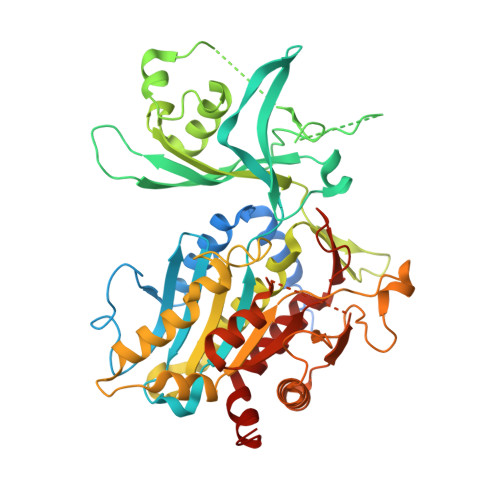
Higher-order assemblies of oligomeric cargo receptor complexes form the membrane scaffold of the Cvt vesicle.

Bertipaglia, C.Schneider, S.Jakobi, A.J.Tarafder, A.K.Bykov, Y.S.Picco, A.Kukulski, W.Kosinski, J.Hagen, W.J.Ravichandran, A.C.Wilmanns, M.Kaksonen, M.Briggs, J.A.Sachse, C.

(2016) EMBO Rep 17: 1044-1060

  • DOI: https://doi.org/10.15252/embr.201541960
  • Primary Citation Related Structures: 
    5JM0, 5JM6, 5JM9

  • PubMed Abstract: 

    Selective autophagy is the mechanism by which large cargos are specifically sequestered for degradation. The structural details of cargo and receptor assembly giving rise to autophagic vesicles remain to be elucidated. We utilize the yeast cytoplasm-to-vacuole targeting (Cvt) pathway, a prototype of selective autophagy, together with a multi-scale analysis approach to study the molecular structure of Cvt vesicles. We report the oligomeric nature of the major Cvt cargo Ape1 with a combined 2.8 Å X-ray and negative stain EM structure, as well as the secondary cargo Ams1 with a 6.3 Å cryo-EM structure. We show that the major dodecameric cargo prApe1 exhibits a tendency to form higher-order chain structures that are broken upon interaction with the receptor Atg19 in vitro The stoichiometry of these cargo-receptor complexes is key to maintaining the size of the Cvt aggregate in vivo Using correlative light and electron microscopy, we further visualize key stages of Cvt vesicle biogenesis. Our findings suggest that Atg19 interaction limits Ape1 aggregate size while serving as a vehicle for vacuolar delivery of tetrameric Ams1.


  • Organizational Affiliation
    • Structural and Computational Biology Unit, European Molecular Biology Laboratory, Heidelberg, Germany.

Macromolecule Content 

  • Total Structure Weight: 57.16 kDa 
  • Atom Count: 3,418 
  • Modeled Residue Count: 438 
  • Deposited Residue Count: 514 
  • Unique protein chains: 1

Macromolecules

Find similar proteins by:|  3D Structure
Entity ID: 1
MoleculeChains  Sequence LengthOrganismDetailsImage
Vacuolar aminopeptidase 1514Saccharomyces cerevisiaeMutation(s): 0 
Gene Names: APE1APILAP4YSC1YKL103CYKL455
EC: 3.4.11.22
UniProt
Find proteins for P14904 (Saccharomyces cerevisiae (strain ATCC 204508 / S288c))
Explore P14904 
Go to UniProtKB:  P14904
Entity Groups
Sequence Clusters30% Identity50% Identity70% Identity90% Identity95% Identity100% Identity
UniProt GroupP14904
Sequence Annotations
Expand
Reference Sequence

Experimental Data & Validation

Experimental Data

  • Method: ELECTRON MICROSCOPY
  • Resolution: 24.0 Å
  • Aggregation State: PARTICLE 
  • Reconstruction Method: SINGLE PARTICLE 
EM Software:
TaskSoftware PackageVersion
RECONSTRUCTIONSPIDER19.09

Structure Validation

View Full Validation Report


Entry History 

Deposition Data

Revision History  (Full details and data files)

  • Version 1.0: 2016-06-15
    Type: Initial release
  • Version 1.1: 2016-06-22
    Changes: Database references
  • Version 1.2: 2016-06-29
    Changes: Database references
  • Version 1.3: 2016-07-13
    Changes: Database references
  • Version 1.4: 2017-08-02
    Changes: Data collection, Refinement description
  • Version 1.5: 2017-08-30
    Changes: Data collection
  • Version 1.6: 2024-05-15
    Changes: Data collection, Database references, Derived calculations, Refinement description