5I69 | pdb_00005i69

MBP-MamC magnetite-interaction component mutant-D70A


Experimental Data Snapshot

  • Method: X-RAY DIFFRACTION
  • Resolution: 2.70 Å
  • R-Value Free: 
    0.293 (Depositor), 0.293 (DCC) 
  • R-Value Work: 
    0.204 (Depositor), 0.213 (DCC) 
  • R-Value Observed: 
    0.209 (Depositor) 

Starting Model: experimental
View more details

wwPDB Validation 3D Report Full Report

Validation slider image for 5I69

This is version 2.1 of the entry. See complete history

Literature

Structure-function studies of the magnetite-biomineralizing magnetosome-associated protein MamC.

Nudelman, H.Valverde-Tercedor, C.Kolusheva, S.Perez Gonzalez, T.Widdrat, M.Grimberg, N.Levi, H.Nelkenbaum, O.Davidov, G.Faivre, D.Jimenez-Lopez, C.Zarivach, R.

(2016) J Struct Biol 194: 244-252

  • DOI: https://doi.org/10.1016/j.jsb.2016.03.001
  • Primary Citation Related Structures: 
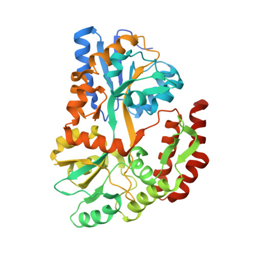
    5I69

  • PubMed Abstract: 

    Magnetotactic bacteria are Gram-negative bacteria that navigate along geomagnetic fields using the magnetosome, an organelle that consists of a membrane-enveloped magnetic nanoparticle. Magnetite formation and its properties are controlled by a specific set of proteins. MamC is a small magnetosome-membrane protein that is known to be active in iron biomineralization but its mechanism has yet to be clarified. Here, we studied the relationship between the MamC magnetite-interaction loop (MIL) structure and its magnetite interaction using an inert biomineralization protein-MamC chimera. Our determined structure shows an alpha-helical fold for MamC-MIL with highly charged surfaces. Additionally, the MamC-MIL induces the formation of larger magnetite crystals compared to protein-free and inert biomineralization protein control experiments. We suggest that the connection between the MamC-MIL structure and the protein's charged surfaces is crucial for magnetite binding and thus for the size control of the magnetite nanoparticles.


  • Organizational Affiliation
    • Department of Life Sciences and the National Institute for Biotechnology in the Negev, Ben-Gurion University of the Negev, Beer Sheva, Israel.

Macromolecule Content 

  • Total Structure Weight: 45.59 kDa 
  • Atom Count: 3,068 
  • Modeled Residue Count: 380 
  • Deposited Residue Count: 412 
  • Unique protein chains: 1

Macromolecules

Find similar proteins by:|  3D Structure
Entity ID: 1
MoleculeChains  Sequence LengthOrganismDetailsImage
Maltose-binding periplasmic protein,Tightly bound bacterial magnetic particle protein,Maltose-binding periplasmic protein412Escherichia coliParamagnetospirillum magneticum AMB-1Mutation(s): 1 
Gene Names: malEb4034JW3994mms13amb0951
UniProt
Find proteins for P0AEX9 (Escherichia coli (strain K12))
Explore P0AEX9 
Go to UniProtKB:  P0AEX9
Find proteins for Q2W8S0 (Paramagnetospirillum magneticum (strain ATCC 700264 / AMB-1))
Explore Q2W8S0 
Go to UniProtKB:  Q2W8S0
Entity Groups
Sequence Clusters30% Identity50% Identity70% Identity90% Identity95% Identity100% Identity
UniProt GroupsQ2W8S0P0AEX9
Sequence Annotations
Expand
Reference Sequence

Oligosaccharides

Help  
Entity ID: 2
MoleculeChains Length2D Diagram GlycosylationD Interactions
alpha-D-glucopyranose-(1-4)-alpha-D-glucopyranose
B
2N/A
Glycosylation Resources
GlyTouCan: G07411ON
GlyCosmos: G07411ON

Small Molecules

Ligands 1 Unique
IDChains Name / Formula / InChI Key2D Diagram3D Interactions
SO4

Query on SO4


Download:Ideal Coordinates CCD File
C [auth A]SULFATE ION
O4 S
QAOWNCQODCNURD-UHFFFAOYSA-L

Biologically Interesting Molecules (External Reference) 

1 Unique

Experimental Data & Validation

Experimental Data

  • Method: X-RAY DIFFRACTION
  • Resolution: 2.70 Å
  • R-Value Free:  0.293 (Depositor), 0.293 (DCC) 
  • R-Value Work:  0.204 (Depositor), 0.213 (DCC) 
  • R-Value Observed: 0.209 (Depositor) 
Space Group: P 43 21 2
Unit Cell:
Length ( Å )Angle ( ˚ )
a = 67.236α = 90
b = 67.236β = 90
c = 209.019γ = 90
Software Package:
Software NamePurpose
REFMACrefinement
HKL-2000data reduction
HKL-2000data scaling
PHASERphasing
Cootmodel building

Structure Validation

View Full Validation Report


Entry History 

Revision History  (Full details and data files)

  • Version 1.0: 2016-03-23
    Type: Initial release
  • Version 1.1: 2016-05-04
    Changes: Database references
  • Version 2.0: 2020-07-29
    Type: Remediation
    Reason: Carbohydrate remediation
    Changes: Atomic model, Data collection, Derived calculations, Non-polymer description, Structure summary
  • Version 2.1: 2024-01-10
    Changes: Data collection, Database references, Refinement description, Structure summary