5I50 | pdb_00005i50

Structure of OmoMYC bound to double-stranded DNA


Experimental Data Snapshot

  • Method: X-RAY DIFFRACTION
  • Resolution: 2.70 Å
  • R-Value Free: 
    0.279 (Depositor), 0.281 (DCC) 
  • R-Value Work: 
    0.227 (Depositor), 0.232 (DCC) 
  • R-Value Observed: 
    0.229 (Depositor) 

Starting Model: experimental
View more details

wwPDB Validation 3D Report Full Report

Validation slider image for 5I50

This is version 1.4 of the entry. See complete history

Literature

OmoMYC blunts promoter invasion by oncogenic MYC to inhibit gene expression characteristic of MYC-dependent tumors.

Jung, L.A.Gebhardt, A.Koelmel, W.Ade, C.P.Walz, S.Kuper, J.von Eyss, B.Letschert, S.Redel, C.d'Artista, L.Biankin, A.Zender, L.Sauer, M.Wolf, E.Evan, G.Kisker, C.Eilers, M.

(2017) Oncogene 36: 1911-1924

  • DOI: https://doi.org/10.1038/onc.2016.354
  • Primary Citation Related Structures: 
    5I4Z, 5I50

  • PubMed Abstract: 

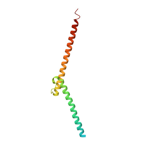
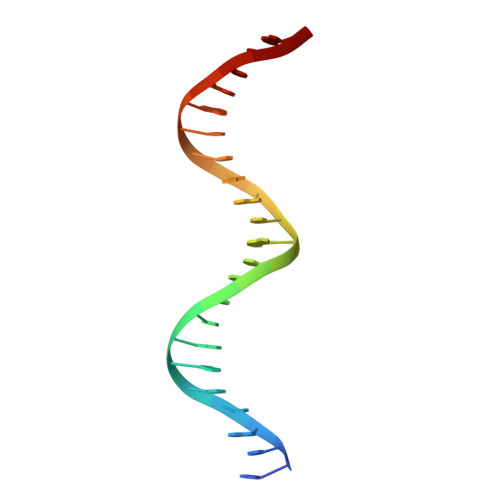
    MYC genes have both essential roles during normal development and exert oncogenic functions during tumorigenesis. Expression of a dominant-negative allele of MYC, termed OmoMYC, can induce rapid tumor regression in mouse models with little toxicity for normal tissues. How OmoMYC discriminates between physiological and oncogenic functions of MYC is unclear. We have solved the crystal structure of OmoMYC and show that it forms a stable homodimer and as such recognizes DNA in the same manner as the MYC/MAX heterodimer. OmoMYC attenuates both MYC-dependent activation and repression by competing with MYC/MAX for binding to chromatin, effectively lowering MYC/MAX occupancy at its cognate binding sites. OmoMYC causes the largest decreases in promoter occupancy and changes in expression on genes that are invaded by oncogenic MYC levels. A signature of OmoMYC-regulated genes defines subgroups with high MYC levels in multiple tumor entities and identifies novel targets for the eradication of MYC-driven tumors.


  • Organizational Affiliation
    • Theodor Boveri Institute, Biocenter, University of Würzburg, Würzburg, Germany.

Macromolecule Content 

  • Total Structure Weight: 41.58 kDa 
  • Atom Count: 2,462 
  • Modeled Residue Count: 232 
  • Deposited Residue Count: 280 
  • Unique protein chains: 1
  • Unique nucleic acid chains: 2

Macromolecules


Find similar proteins by:|  3D Structure
Entity ID: 1
MoleculeChains  Sequence LengthOrganismDetailsImage
Myc proto-oncogene protein
A, B
118Homo sapiensMutation(s): 4 
Gene Names: MYCBHLHE39
UniProt & NIH Common Fund Data Resources
Find proteins for P01106 (Homo sapiens)
Explore P01106 
Go to UniProtKB:  P01106
PHAROS:  P01106
GTEx:  ENSG00000136997 
Entity Groups
Sequence Clusters30% Identity50% Identity70% Identity90% Identity95% Identity100% Identity
UniProt GroupP01106
Sequence Annotations
Expand
Reference Sequence
Find similar nucleic acids by:  Sequence
Entity ID: 2
MoleculeChains LengthOrganismImage
DNA (5'-D(P*CP*AP*CP*CP*CP*GP*GP*TP*CP*AP*CP*GP*TP*GP*GP*CP*CP*TP*AP*CP*AP*C)-3')22synthetic construct
Sequence Annotations
Expand
Reference Sequence
Find similar nucleic acids by:  Sequence
Entity ID: 3
MoleculeChains LengthOrganismImage
DNA (5'-D(P*GP*TP*GP*TP*AP*GP*GP*CP*CP*AP*CP*GP*TP*GP*AP*CP*CP*GP*GP*GP*TP*G)-3')22synthetic construct
Sequence Annotations
Expand
Reference Sequence

Experimental Data & Validation

Experimental Data

  • Method: X-RAY DIFFRACTION
  • Resolution: 2.70 Å
  • R-Value Free:  0.279 (Depositor), 0.281 (DCC) 
  • R-Value Work:  0.227 (Depositor), 0.232 (DCC) 
  • R-Value Observed: 0.229 (Depositor) 
Space Group: P 1 21 1
Unit Cell:
Length ( Å )Angle ( ˚ )
a = 36.538α = 90
b = 95.545β = 105.9
c = 64.944γ = 90
Software Package:
Software NamePurpose
PHENIXrefinement
XDSdata reduction
XDSdata scaling
PHASERphasing

Structure Validation

View Full Validation Report


Entry History 

& Funding Information

Deposition Data


Funding OrganizationLocationGrant Number
German Research FoundationGermany2341

Revision History  (Full details and data files)

  • Version 1.0: 2016-10-26
    Type: Initial release
  • Version 1.1: 2017-04-19
    Changes: Database references
  • Version 1.2: 2019-02-20
    Changes: Author supporting evidence, Data collection, Derived calculations, Source and taxonomy
  • Version 1.3: 2024-01-10
    Changes: Data collection, Database references, Refinement description
  • Version 1.4: 2024-11-13
    Changes: Structure summary