5HQ4 | pdb_00005hq4

A Glycoside Hydrolase Family 97 enzyme from Pseudoalteromonas sp. strain K8


Experimental Data Snapshot

  • Method: X-RAY DIFFRACTION
  • Resolution: 1.93 Å
  • R-Value Free: 
    0.160 (Depositor), 0.161 (DCC) 
  • R-Value Work: 
    0.144 (Depositor), 0.145 (DCC) 
  • R-Value Observed: 
    0.144 (Depositor) 

Starting Model: experimental
View more details

wwPDB Validation 3D Report Full Report

Validation slider image for 5HQ4

This is version 1.1 of the entry. See complete history

Literature

Structures of PspAG97A alpha-glucoside hydrolase reveal a novel mechanism for chloride induced activation.

He, C.Li, J.Li, W.Xue, Y.Fang, Z.Fang, W.Zhang, X.Wang, X.Xiao, Y.

(2016) J Struct Biol 196: 426-436

  • DOI: https://doi.org/10.1016/j.jsb.2016.09.009
  • Primary Citation Related Structures: 
    5HQ4, 5HQA, 5HQB, 5HQC

  • PubMed Abstract: 

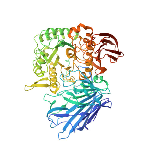
    Here we report the first crystal structure of a secretory α-glucoside hydrolase isolated from Pseudoalteromonas sp. K8, PspAG97A, which belongs to glycoside hydrolase family 97 and exhibits halophilic property. PspAG97A lacks an acidic surface, that is considered essential for protein stability at high salinity. Interestingly, PspAG97A unusually contains a chloride ion coordinated by the guanidinium group of Arg171 and the main chain amide groups of Tyr172 and Glu173 at the active site. The structures of PspAG97A complexed with acarbose and panose demonstrate that residues Glu173, Arg171 and Asn170 for subsite +1 decide the substrate specificity of the enzyme for the α-1,6-glucosidic linkage. Structural alterations observed in the R171K variant and enzyme kinetic experiments focusing on chloride assisted activation suggest that the active site chloride serves to properly orient Glu173, Arg171 and Asn170 to facilitate substrate recognition. Furthermore, the chloride assists the binding of Glu173 to the conserved calcium ion and plays an essential role in properly positioning the base catalyst Glu456. In sum, our results provide valuable insight into the structural basis of protein halophilicity.


  • Organizational Affiliation
    • School of Life Sciences, Anhui University, Hefei, Anhui 230601, China; Anhui Provincial Engineering Technology Research Center of Microorganisms and Biocatalysis, Hefei, Anhui 230601, China.

Macromolecule Content 

  • Total Structure Weight: 76.31 kDa 
  • Atom Count: 6,311 
  • Modeled Residue Count: 662 
  • Deposited Residue Count: 669 
  • Unique protein chains: 1

Macromolecules

Find similar proteins by:|  3D Structure
Entity ID: 1
MoleculeChains  Sequence LengthOrganismDetailsImage
Alpha-glucosidase669Pseudoalteromonas sp. K8Mutation(s): 0 
UniProt
Find proteins for A0A0Y0DFX2 (Pseudoalteromonas sp. K8)
Explore A0A0Y0DFX2 
Go to UniProtKB:  A0A0Y0DFX2
Entity Groups
Sequence Clusters30% Identity50% Identity70% Identity90% Identity95% Identity100% Identity
UniProt GroupA0A0Y0DFX2
Sequence Annotations
Expand
Reference Sequence

Small Molecules

Ligands 6 Unique
IDChains Name / Formula / InChI Key2D Diagram3D Interactions
GOL

Query on GOL


Download:Ideal Coordinates CCD File
B [auth A],
C [auth A],
D [auth A],
E [auth A],
F [auth A]
GLYCEROL
C3 H8 O3
PEDCQBHIVMGVHV-UHFFFAOYSA-N
FMT

Query on FMT


Download:Ideal Coordinates CCD File
K [auth A],
L [auth A],
M [auth A]
FORMIC ACID
C H2 O2
BDAGIHXWWSANSR-UHFFFAOYSA-N
CA

Query on CA


Download:Ideal Coordinates CCD File
G [auth A]CALCIUM ION
Ca
BHPQYMZQTOCNFJ-UHFFFAOYSA-N
CL

Query on CL


Download:Ideal Coordinates CCD File
H [auth A],
I [auth A]
CHLORIDE ION
Cl
VEXZGXHMUGYJMC-UHFFFAOYSA-M
MG

Query on MG


Download:Ideal Coordinates CCD File
J [auth A]MAGNESIUM ION
Mg
JLVVSXFLKOJNIY-UHFFFAOYSA-N
UNX

Query on UNX


Download:Ideal Coordinates CCD File
N [auth A]UNKNOWN ATOM OR ION
X

Experimental Data & Validation

Experimental Data

  • Method: X-RAY DIFFRACTION
  • Resolution: 1.93 Å
  • R-Value Free:  0.160 (Depositor), 0.161 (DCC) 
  • R-Value Work:  0.144 (Depositor), 0.145 (DCC) 
  • R-Value Observed: 0.144 (Depositor) 
Space Group: P 32 2 1
Unit Cell:
Length ( Å )Angle ( ˚ )
a = 119.545α = 90
b = 119.545β = 90
c = 182.757γ = 120
Software Package:
Software NamePurpose
PHENIXrefinement
HKL-2000data collection
HKL-2000data scaling
PDB_EXTRACTdata extraction
HKL-2000data reduction
PHASERphasing

Structure Validation

View Full Validation Report


Entry History 

Deposition Data

Revision History  (Full details and data files)

  • Version 1.0: 2017-01-11
    Type: Initial release
  • Version 1.1: 2023-11-08
    Changes: Data collection, Database references, Derived calculations, Refinement description