5H6B | pdb_00005h6b

Crystal structure of a thermostable lipase from Marine Streptomyces


Experimental Data Snapshot

  • Method: X-RAY DIFFRACTION
  • Resolution: 2.30 Å
  • R-Value Free: 
    0.206 (Depositor), 0.212 (DCC) 
  • R-Value Work: 
    0.178 (Depositor), 0.189 (DCC) 
  • R-Value Observed: 
    0.180 (Depositor) 

wwPDB Validation 3D Report Full Report

Validation slider image for 5H6B

This is version 1.3 of the entry. See complete history

Literature

Crystal structure of a lipase from Streptomyces sp. strain W007 - implications for thermostability and regiospecificity

Zhao, Z.Hou, S.Lan, D.Wang, X.Liu, J.Khan, F.I.Wang, Y.

(2017) FEBS J 284: 3506-3519

  • DOI: https://doi.org/10.1111/febs.14211
  • Primary Citation Related Structures: 
    5H6B, 5H6G

  • PubMed Abstract: 

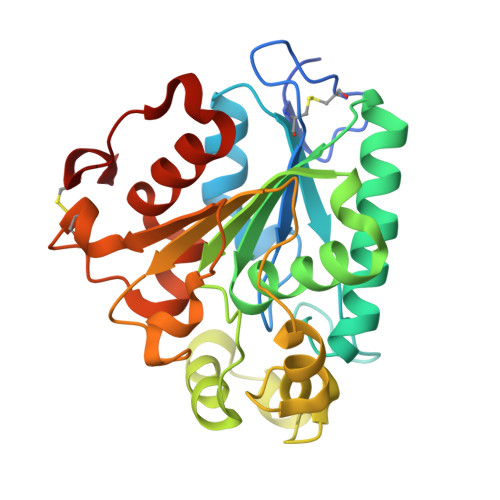
    MAS1 from marine Streptomyces sp. strain W007 belongs to the bacterial lipase I.7 subfamily and is characterized as a thermostable and nonregiospecific lipase. To shed light on the catalytic mechanism of MAS1, we determined its crystal structure with closed conformation in two crystal forms at 2.3 Å resolution. MAS1 adopts the canonical α/β hydrolase core fold with its catalytic triad being formed by S109, D200 and H232. Structural analysis and biochemical assays revealed that disulfide bonds and salt bridges play a vital role in the thermostability of MAS1. In addition, we discovered that the replacement of H108 with a tryptophan converts MAS1 from a nonregiospecific to an sn-1,3-specific lipase, suggesting the functional importance of the second position from the conserved pentapeptide motif in defining the regiospecificity of MAS1. Our present study provides insights into the molecular basis for the thermostability and regiospecificity of MAS1, and it may aid in the rational design of thermostable or regiospecific lipases for potential industrial applications. Structural data are available in the Protein Data Bank database under the accession numbers 5H6B and 5H6G.


  • Organizational Affiliation
    • School of Bioscience and Bioengineering, South China University of Technology, Guangzhou, China.

Macromolecule Content 

  • Total Structure Weight: 28.58 kDa 
  • Atom Count: 1,970 
  • Modeled Residue Count: 252 
  • Deposited Residue Count: 265 
  • Unique protein chains: 1

Macromolecules

Find similar proteins by:|  3D Structure
Entity ID: 1
MoleculeChains  Sequence LengthOrganismDetailsImage
Putative secreted lipase265Streptomyces sp. W007Mutation(s): 0 
Gene Names: SPW_1544
EC: 3.1.1.3

Small Molecules

Ligands 3 Unique
IDChains Name / Formula / InChI Key2D Diagram3D Interactions
IMD

Query on IMD


Download:Ideal Coordinates CCD File
H [auth A],
I [auth A]
IMIDAZOLE
C3 H5 N2
RAXXELZNTBOGNW-UHFFFAOYSA-O
ZN

Query on ZN


Download:Ideal Coordinates CCD File
B [auth A],
C [auth A],
D [auth A],
E [auth A]
ZINC ION
Zn
PTFCDOFLOPIGGS-UHFFFAOYSA-N
ACT

Query on ACT


Download:Ideal Coordinates CCD File
F [auth A],
G [auth A]
ACETATE ION
C2 H3 O2
QTBSBXVTEAMEQO-UHFFFAOYSA-M

Experimental Data & Validation

Experimental Data

  • Method: X-RAY DIFFRACTION
  • Resolution: 2.30 Å
  • R-Value Free:  0.206 (Depositor), 0.212 (DCC) 
  • R-Value Work:  0.178 (Depositor), 0.189 (DCC) 
  • R-Value Observed: 0.180 (Depositor) 
Space Group: P 64
Unit Cell:
Length ( Å )Angle ( ˚ )
a = 90.73α = 90
b = 90.73β = 90
c = 70.62γ = 120
Software Package:
Software NamePurpose
MOSFLMdata collection
Aimlessdata scaling
PHASESphasing
REFMACrefinement
PDB_EXTRACTdata extraction
MOSFLMdata reduction

Structure Validation

View Full Validation Report


Entry History 

Deposition Data

Revision History  (Full details and data files)

  • Version 1.0: 2017-09-20
    Type: Initial release
  • Version 1.1: 2017-10-18
    Changes: Database references
  • Version 1.2: 2017-11-01
    Changes: Database references
  • Version 1.3: 2024-11-20
    Changes: Data collection, Database references, Derived calculations, Structure summary