5FRQ | pdb_00005frq

Crystal Structure of Helicobacter pylori beta clamp bound to DNA ligase peptide


Experimental Data Snapshot

  • Method: X-RAY DIFFRACTION
  • Resolution: 2.90 Å
  • R-Value Free: 
    0.262 (Depositor) 
  • R-Value Work: 
    0.231 (Depositor) 
  • R-Value Observed: 
    0.233 (Depositor) 

Starting Model: experimental
View more details

wwPDB Validation 3D Report Full Report

Validation slider image for 5FRQ

This is version 1.2 of the entry. See complete history

Literature

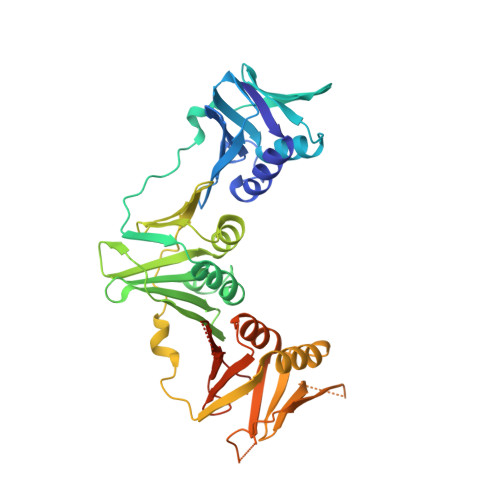
Structural Insight Into Beta-Clamp and its Interaction with DNA Ligase in Helicobacter Pylori

Pandey, P.Tarique, K.F.Mazumder, M.Abdul Rehman, S.A.Gourinath, S.

(2016) Sci Rep 6: 31181

  • DOI: https://doi.org/10.1038/srep31181
  • Primary Citation Related Structures: 
    4RKI, 4S3I, 5FRQ

  • PubMed Abstract: 

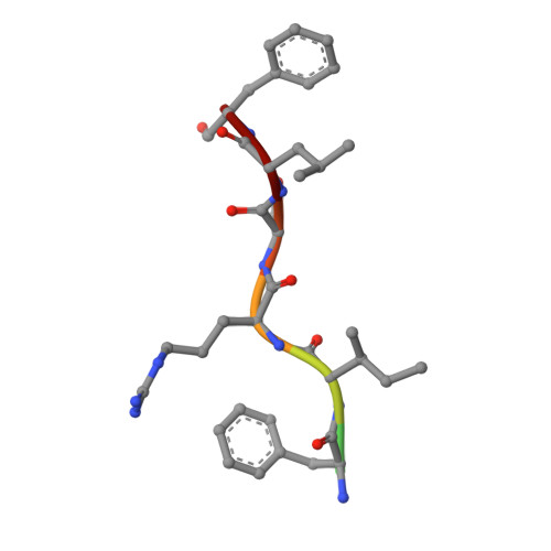
    Helicobacter pylori, a gram-negative and microaerophilic bacterium, is the major cause of chronic gastritis, gastric ulcers and gastric cancer. Owing to its central role, DNA replication machinery has emerged as a prime target for the development of antimicrobial drugs. Here, we report 2Å structure of β-clamp from H. pylori (Hpβ-clamp), which is one of the critical components of DNA polymerase III. Despite of similarity in the overall fold of eubacterial β-clamp structures, some distinct features in DNA interacting loops exists that have not been reported previously. The in silico prediction identified the potential binders of β-clamp such as alpha subunit of DNA pol III and DNA ligase with identification of β-clamp binding regions in them and validated by SPR studies. Hpβ-clamp interacts with DNA ligase in micromolar binding affinity. Moreover, we have successfully determined the co-crystal structure of β-clamp with peptide from DNA ligase (not reported earlier in prokaryotes) revealing the region from ligase that interacts with β-clamp.


  • Organizational Affiliation
    • School of Life Sciences, Jawaharlal Nehru University, New Delhi, India.

Macromolecule Content 

  • Total Structure Weight: 176.13 kDa 
  • Atom Count: 11,507 
  • Modeled Residue Count: 1,449 
  • Deposited Residue Count: 1,552 
  • Unique protein chains: 2

Macromolecules

Find similar proteins by:|  3D Structure
Entity ID: 1
MoleculeChains  Sequence LengthOrganismDetailsImage
DNA POLYMERASE III SUBUNIT BETA
A, B, C, D
384Helicobacter pylori 26695Mutation(s): 0 
EC: 2.7.7.7
UniProt
Find proteins for O25242 (Helicobacter pylori (strain ATCC 700392 / 26695))
Explore O25242 
Go to UniProtKB:  O25242
Entity Groups
Sequence Clusters30% Identity50% Identity70% Identity90% Identity95% Identity100% Identity
UniProt GroupO25242
Sequence Annotations
Expand
Reference Sequence
Find similar proteins by:  Sequence   |   3D Structure  
Entity ID: 2
MoleculeChains  Sequence LengthOrganismDetailsImage
DNA LIGASEE [auth G],
F [auth L]
8Helicobacter pylori 26695Mutation(s): 0 
EC: 6.5.1.2
UniProt
Find proteins for O25336 (Helicobacter pylori (strain ATCC 700392 / 26695))
Explore O25336 
Go to UniProtKB:  O25336
Entity Groups
UniProt GroupO25336
Sequence Annotations
Expand
Reference Sequence

Experimental Data & Validation

Experimental Data

  • Method: X-RAY DIFFRACTION
  • Resolution: 2.90 Å
  • R-Value Free:  0.262 (Depositor) 
  • R-Value Work:  0.231 (Depositor) 
  • R-Value Observed: 0.233 (Depositor) 
Space Group: P 21 21 21
Unit Cell:
Length ( Å )Angle ( ˚ )
a = 64.829α = 90
b = 146.147β = 90
c = 179.075γ = 90
Software Package:
Software NamePurpose
REFMACrefinement

Structure Validation

View Full Validation Report


Entry History 

Deposition Data

Revision History  (Full details and data files)

  • Version 1.0: 2016-08-17
    Type: Initial release
  • Version 1.1: 2019-12-11
    Changes: Derived calculations, Other
  • Version 1.2: 2024-01-10
    Changes: Data collection, Database references, Refinement description