5F6I | pdb_00005f6i

Crystal Structure of Tier 2 Neutralizing Antibody DH428 from a Rhesus Macaque


Experimental Data Snapshot

  • Method: X-RAY DIFFRACTION
  • Resolution: 2.32 Å
  • R-Value Free: 
    0.265 (Depositor), 0.271 (DCC) 
  • R-Value Work: 
    0.218 (Depositor), 0.227 (DCC) 
  • R-Value Observed: 
    0.220 (Depositor) 

Starting Model: experimental
View more details

wwPDB Validation 3D Report Full Report

Validation slider image for 5F6I

This is version 1.4 of the entry. See complete history

Literature

Structural Constraints of Vaccine-Induced Tier-2 Autologous HIV Neutralizing Antibodies Targeting the Receptor-Binding Site.

Bradley, T.Fera, D.Bhiman, J.Eslamizar, L.Lu, X.Anasti, K.Zhang, R.Sutherland, L.L.Scearce, R.M.Bowman, C.M.Stolarchuk, C.Lloyd, K.E.Parks, R.Eaton, A.Foulger, A.Nie, X.Karim, S.S.Barnett, S.Kelsoe, G.Kepler, T.B.Alam, S.M.Montefiori, D.C.Moody, M.A.Liao, H.X.Morris, L.Santra, S.Harrison, S.C.Haynes, B.F.

(2016) Cell Rep 14: 43-54

  • DOI: https://doi.org/10.1016/j.celrep.2015.12.017
  • Primary Citation Related Structures: 
    5F6H, 5F6I, 5F6J

  • PubMed Abstract: 

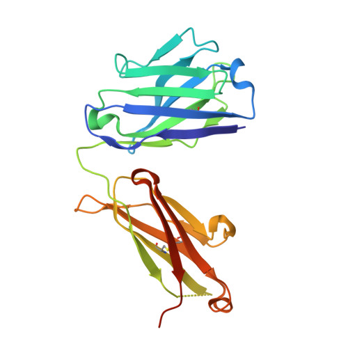
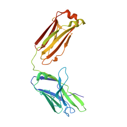
    Antibodies that neutralize autologous transmitted/founder (TF) HIV occur in most HIV-infected individuals and can evolve to neutralization breadth. Autologous neutralizing antibodies (nAbs) against neutralization-resistant (Tier-2) viruses are rarely induced by vaccination. Whereas broadly neutralizing antibody (bnAb)-HIV-Envelope structures have been defined, the structures of autologous nAbs have not. Here, we show that immunization with TF mutant Envs gp140 oligomers induced high-titer, V5-dependent plasma neutralization for a Tier-2 autologous TF evolved mutant virus. Structural analysis of autologous nAb DH427 revealed binding to V5, demonstrating the source of narrow nAb specificity and explaining the failure to acquire breadth. Thus, oligomeric TF Envs can elicit autologous nAbs to Tier-2 HIVs, but induction of bnAbs will require targeting of precursors of B cell lineages that can mature to heterologous neutralization.


  • Organizational Affiliation
    • Duke Human Vaccine Institute, Departments of Medicine, Surgery and Immunology, Duke University School of Medicine, Durham, NC 27710, USA. Electronic address: todd.bradley@duke.edu.

Macromolecule Content 

  • Total Structure Weight: 47.22 kDa 
  • Atom Count: 3,225 
  • Modeled Residue Count: 420 
  • Deposited Residue Count: 443 
  • Unique protein chains: 2

Macromolecules

Find similar proteins by:|  3D Structure
Entity ID: 1
MoleculeChains  Sequence LengthOrganismDetailsImage
DH428 Antibody Heavy ChainA [auth B]227Macaca mulattaMutation(s): 0 
Find similar proteins by:|  3D Structure
Entity ID: 2
MoleculeChains  Sequence LengthOrganismDetailsImage
DH428 Antibody Light ChainB [auth C]216Macaca mulattaMutation(s): 0 

Experimental Data & Validation

Experimental Data

  • Method: X-RAY DIFFRACTION
  • Resolution: 2.32 Å
  • R-Value Free:  0.265 (Depositor), 0.271 (DCC) 
  • R-Value Work:  0.218 (Depositor), 0.227 (DCC) 
  • R-Value Observed: 0.220 (Depositor) 
Space Group: P 21 2 21
Unit Cell:
Length ( Å )Angle ( ˚ )
a = 73.106α = 90
b = 74.628β = 90
c = 102.62γ = 90
Software Package:
Software NamePurpose
PHENIXrefinement
PDB_EXTRACTdata extraction
Cootmodel building
XSCALEdata scaling
XDSdata reduction
PHASERphasing

Structure Validation

View Full Validation Report


Entry History 

Deposition Data

Revision History  (Full details and data files)

  • Version 1.0: 2016-01-13
    Type: Initial release
  • Version 1.1: 2016-01-20
    Changes: Database references
  • Version 1.2: 2017-07-05
    Changes: Source and taxonomy
  • Version 1.3: 2023-09-27
    Changes: Data collection, Database references, Refinement description
  • Version 1.4: 2024-10-23
    Changes: Structure summary