5E63

K262A mutant of I-SmaMI


Experimental Data Snapshot

  • Method: X-RAY DIFFRACTION
  • Resolution: 2.60 Å
  • R-Value Free: 0.279 
  • R-Value Work: 0.189 
  • R-Value Observed: 0.193 

Starting Model: experimental
View more details

wwPDB Validation   3D Report Full Report


This is version 1.3 of the entry. See complete history


Literature

The Structural Basis of Asymmetry in DNA Binding and Cleavage as Exhibited by the I-SmaMI LAGLIDADG Meganuclease.

Shen, B.W.Lambert, A.Walker, B.C.Stoddard, B.L.Kaiser, B.K.

(2016) J Mol Biol 428: 206-220

  • DOI: https://doi.org/10.1016/j.jmb.2015.12.005
  • Primary Citation of Related Structures:  
    5E5O, 5E5P, 5E5S, 5E63, 5E67

  • PubMed Abstract: 

    LAGLIDADG homing endonucleases ("meganucleases") are highly specific DNA cleaving enzymes that are used for genome engineering. Like other enzymes that act on DNA targets, meganucleases often display binding affinities and cleavage activities that are dominated by one protein domain. To decipher the underlying mechanism of asymmetric DNA recognition and catalysis, we identified and characterized a new monomeric meganuclease (I-SmaMI), which belongs to a superfamily of homologous enzymes that recognize divergent DNA sequences. We solved a series of crystal structures of the enzyme-DNA complex representing a progression of sequential reaction states, and we compared the structural rearrangements and surface potential distributions within each protein domain against their relative contribution to binding affinity. We then determined the effects of equivalent point mutations in each of the two enzyme active sites to determine whether asymmetry in DNA recognition is translated into corresponding asymmetry in DNA cleavage activity. These experiments demonstrate the structural basis for "dominance" by one protein domain over the other and provide insights into this enzyme's conformational switch from a nonspecific search mode to a more specific recognition mode.


  • Organizational Affiliation

    Division of Basic Sciences, Fred Hutchinson Cancer Research Center, 1100 Fairview Avenue North, Seattle, WA 98109, USA. Electronic address: bshen@fredhutch.org.


Macromolecules

Find similar proteins by:  (by identity cutoff)  |  3D Structure
Entity ID: 1
MoleculeChains Sequence LengthOrganismDetailsImage
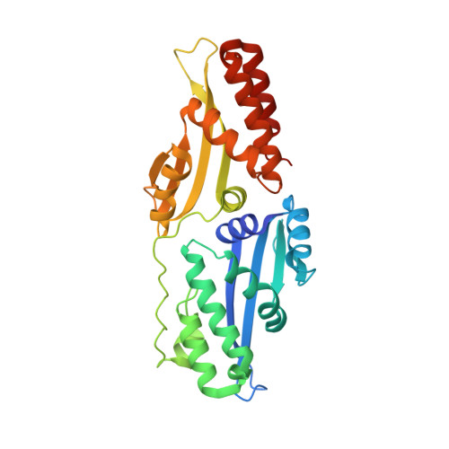
I-SmaMI LAGLIDADG meganuclease302Sordaria macrospora k-hellMutation(s): 1 
Gene Names: SMAC_12671
UniProt
Find proteins for F7WD42 (Sordaria macrospora (strain ATCC MYA-333 / DSM 997 / K(L3346) / K-hell))
Explore F7WD42 
Go to UniProtKB:  F7WD42
Entity Groups  
Sequence Clusters30% Identity50% Identity70% Identity90% Identity95% Identity100% Identity
UniProt GroupF7WD42
Sequence Annotations
Expand
  • Reference Sequence

Find similar nucleic acids by:  Sequence   |   3D Structure  

Entity ID: 2
MoleculeChains LengthOrganismImage
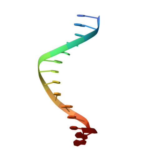
DNA right half site Bottom strand14synthetic construct
Sequence Annotations
Expand
  • Reference Sequence

Find similar nucleic acids by:  Sequence   |   3D Structure  

Entity ID: 3
MoleculeChains LengthOrganismImage
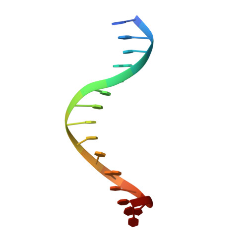
DNA left half site bottom strand11synthetic construct
Sequence Annotations
Expand
  • Reference Sequence

Find similar nucleic acids by:  Sequence   |   3D Structure  

Entity ID: 4
MoleculeChains LengthOrganismImage
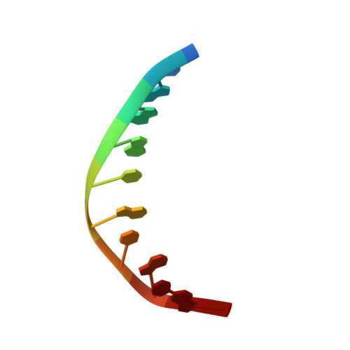
DNA left site top strand15synthetic construct
Sequence Annotations
Expand
  • Reference Sequence

Find similar nucleic acids by:  Sequence   |   3D Structure  

Entity ID: 5
MoleculeChains LengthOrganismImage
DNA (5'-D(P*CP*AP*GP*GP*TP*GP*TP*AP*CP*G)-3')10synthetic construct
Sequence Annotations
Expand
  • Reference Sequence
Experimental Data & Validation

Experimental Data

  • Method: X-RAY DIFFRACTION
  • Resolution: 2.60 Å
  • R-Value Free: 0.279 
  • R-Value Work: 0.189 
  • R-Value Observed: 0.193 
  • Space Group: P 21 21 21
Unit Cell:
Length ( Å )Angle ( ˚ )
a = 59.835α = 90
b = 67.694β = 90
c = 97.78γ = 90
Software Package:
Software NamePurpose
REFMACrefinement
HKL-2000data reduction
SCALEPACKdata scaling
PHASERphasing

Structure Validation

View Full Validation Report


Entry History 

Deposition Data

Revision History  (Full details and data files)

  • Version 1.0: 2016-01-13
    Type: Initial release
  • Version 1.1: 2016-02-10
    Changes: Database references
  • Version 1.2: 2016-04-06
    Changes: Source and taxonomy
  • Version 1.3: 2023-09-27
    Changes: Data collection, Database references, Derived calculations, Refinement description