5D5V

Crystal structure of human Hsf1 with Satellite III repeat DNA


Experimental Data Snapshot

  • Method: X-RAY DIFFRACTION
  • Resolution: 2.55 Å
  • R-Value Free: 0.234 
  • R-Value Work: 0.190 
  • R-Value Observed: 0.192 

Starting Model: experimental
View more details

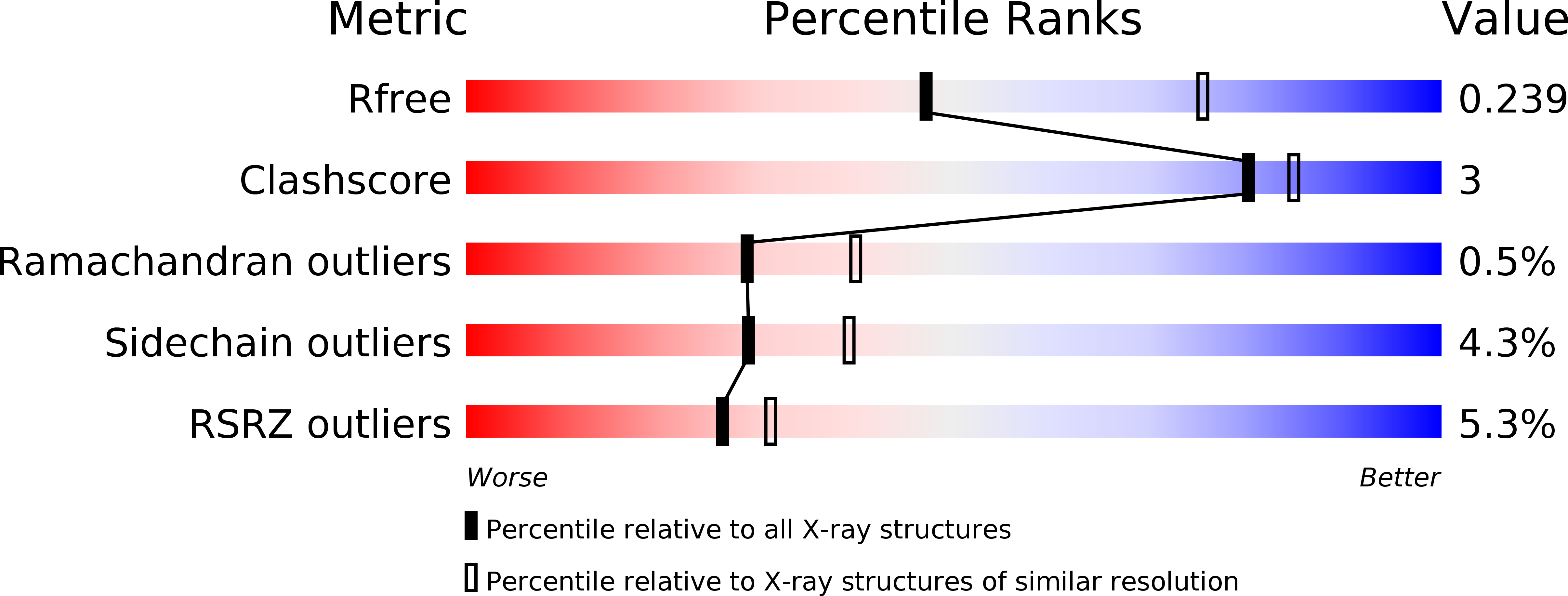
wwPDB Validation   3D Report Full Report


This is version 1.4 of the entry. See complete history


Literature

Structure of human heat-shock transcription factor 1 in complex with DNA.

Neudegger, T.Verghese, J.Hayer-Hartl, M.Hartl, F.U.Bracher, A.

(2016) Nat Struct Mol Biol 23: 140-146

  • DOI: https://doi.org/10.1038/nsmb.3149
  • Primary Citation of Related Structures:  
    5D5U, 5D5V, 5D5W, 5D5X, 5D5Y, 5D5Z, 5D60

  • PubMed Abstract: 

    Heat-shock transcription factor 1 (HSF1) has a central role in mediating the protective response to protein conformational stresses in eukaryotes. HSF1 consists of an N-terminal DNA-binding domain (DBD), a coiled-coil oligomerization domain, a regulatory domain and a transactivation domain. Upon stress, HSF1 trimerizes via its coiled-coil domain and binds to the promoters of heat shock protein-encoding genes. Here, we present cocrystal structures of the human HSF1 DBD in complex with cognate DNA. A comparative analysis of the HSF1 paralog Skn7 from Chaetomium thermophilum showed that single amino acid changes in the DBD can switch DNA binding specificity, thus revealing the structural basis for the interaction of HSF1 with cognate DNA. We used a crystal structure of the coiled-coil domain of C. thermophilum Skn7 to develop a model of the active human HSF1 trimer in which HSF1 embraces the heat-shock-element DNA.


  • Organizational Affiliation

    Department of Cellular Biochemistry, Max Planck Institute of Biochemistry, Martinsried, Germany.


Macromolecules

Find similar proteins by:  (by identity cutoff)  |  3D Structure
Entity ID: 1
MoleculeChains Sequence LengthOrganismDetailsImage
Heat shock factor protein 1A [auth B],
B [auth D]
131Homo sapiensMutation(s): 0 
Gene Names: HSF1HSTF1
UniProt & NIH Common Fund Data Resources
Find proteins for Q00613 (Homo sapiens)
Explore Q00613 
Go to UniProtKB:  Q00613
PHAROS:  Q00613
GTEx:  ENSG00000185122 
Entity Groups  
Sequence Clusters30% Identity50% Identity70% Identity90% Identity95% Identity100% Identity
UniProt GroupQ00613
Sequence Annotations
Expand
  • Reference Sequence

Find similar nucleic acids by:  Sequence   |   3D Structure  

Entity ID: 2
MoleculeChains LengthOrganismImage
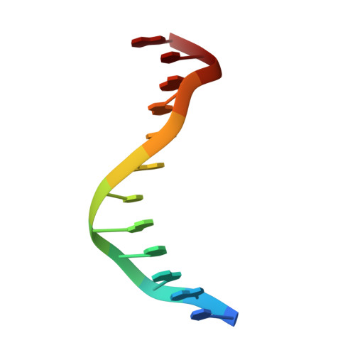
DNAC [auth A]12Homo sapiens
Sequence Annotations
Expand
  • Reference Sequence

Find similar nucleic acids by:  Sequence   |   3D Structure  

Entity ID: 3
MoleculeChains LengthOrganismImage
DNAD [auth C]12Homo sapiens
Sequence Annotations
Expand
  • Reference Sequence
Experimental Data & Validation

Experimental Data

  • Method: X-RAY DIFFRACTION
  • Resolution: 2.55 Å
  • R-Value Free: 0.234 
  • R-Value Work: 0.190 
  • R-Value Observed: 0.192 
  • Space Group: C 1 2 1
Unit Cell:
Length ( Å )Angle ( ˚ )
a = 104.813α = 90
b = 112.678β = 92.56
c = 39.865γ = 90
Software Package:
Software NamePurpose
Aimlessdata scaling
MOLREPphasing
REFMACrefinement
PDB_EXTRACTdata extraction
XDSdata reduction

Structure Validation

View Full Validation Report


Entry History 

Deposition Data

Revision History  (Full details and data files)

  • Version 1.0: 2015-12-30
    Type: Initial release
  • Version 1.1: 2016-01-13
    Changes: Database references
  • Version 1.2: 2016-01-20
    Changes: Structure summary
  • Version 1.3: 2016-02-10
    Changes: Database references
  • Version 1.4: 2024-01-10
    Changes: Data collection, Database references, Refinement description