5D3G | pdb_00005d3g

Structure of HIV-1 Reverse Transcriptase Bound to a Novel 38-mer Hairpin Template-Primer DNA Aptamer


Experimental Data Snapshot

  • Method: X-RAY DIFFRACTION
  • Resolution: 2.30 Å
  • R-Value Free: 
    0.225 (Depositor), 0.228 (DCC) 
  • R-Value Work: 
    0.191 (Depositor), 0.199 (DCC) 
  • R-Value Observed: 
    0.191 (Depositor) 

Starting Model: experimental
View more details

wwPDB Validation 3D Report Full Report

Validation slider image for 5D3G

This is version 3.1 of the entry. See complete history

Literature

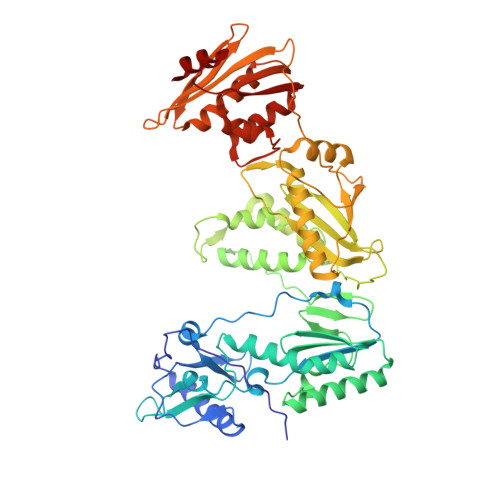
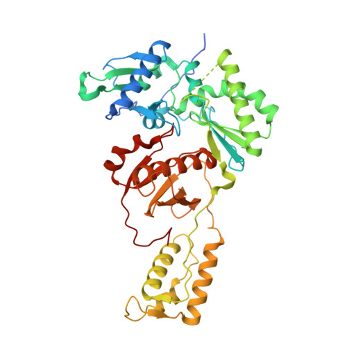
Structure of HIV-1 reverse transcriptase bound to a novel 38-mer hairpin template-primer DNA aptamer.

Miller, M.T.Tuske, S.Das, K.DeStefano, J.J.Arnold, E.

(2016) Protein Sci 25: 46-55

  • DOI: https://doi.org/10.1002/pro.2776
  • Primary Citation Related Structures: 
    5D3G

  • PubMed Abstract: 

    The development of a modified DNA aptamer that binds HIV-1 reverse transcriptase (RT) with ultra-high affinity has enabled the X-ray structure determination of an HIV-1 RT-DNA complex to 2.3 Å resolution without the need for an antibody Fab fragment or RT-DNA cross-linking. The 38-mer hairpin-DNA aptamer has a 15 base-pair duplex, a three-deoxythymidine hairpin loop, and a five-nucleotide 5'-overhang. The aptamer binds RT in a template-primer configuration with the 3'-end positioned at the polymerase active site and has 2'-O-methyl modifications at the second and fourth duplex template nucleotides that interact with the p66 fingers and palm subdomains. This structure represents the highest resolution RT-nucleic acid structure to date. The RT-aptamer complex is catalytically active and can serve as a platform for studying fundamental RT mechanisms and for development of anti-HIV inhibitors through fragment screening and other approaches. Additionally, the structure allows for a detailed look at a unique aptamer design and provides the molecular basis for its remarkably high affinity for RT.


  • Organizational Affiliation
    • Center for Advanced Biotechnology and Medicine, Department of Chemistry and Chemical Biology, Rutgers University, Piscataway, New Jersey, 08854.

Macromolecule Content 

  • Total Structure Weight: 256.99 kDa 
  • Atom Count: 17,868 
  • Modeled Residue Count: 2,003 
  • Deposited Residue Count: 2,074 
  • Unique protein chains: 2
  • Unique nucleic acid chains: 1

Macromolecules


Find similar proteins by:|  3D Structure
Entity ID: 1
MoleculeChains  Sequence LengthOrganismDetailsImage
HIV-1 REVERSE TRANSCRIPTASE P66 subunit
A, C
555Human immunodeficiency virus type 1 BH10Mutation(s): 1 
Gene Names: gag-pol
EC: 2.7.7.49
UniProt
Find proteins for P03366 (Human immunodeficiency virus type 1 group M subtype B (isolate BH10))
Explore P03366 
Go to UniProtKB:  P03366
Entity Groups
Sequence Clusters30% Identity50% Identity70% Identity90% Identity95% Identity100% Identity
UniProt GroupP03366
Sequence Annotations
Expand
Reference Sequence
Find similar proteins by:|  3D Structure
Entity ID: 2
MoleculeChains  Sequence LengthOrganismDetailsImage
HIV-1 REVERSE TRANSCRIPTASE P51 subunit
B, D
444Human immunodeficiency virus type 1 BH10Mutation(s): 1 
Gene Names: gag-pol
EC: 2.7.7.49
UniProt
Find proteins for P03366 (Human immunodeficiency virus type 1 group M subtype B (isolate BH10))
Explore P03366 
Go to UniProtKB:  P03366
Entity Groups
Sequence Clusters30% Identity50% Identity70% Identity90% Identity95% Identity100% Identity
UniProt GroupP03366
Sequence Annotations
Expand
Reference Sequence
Find similar nucleic acids by:  (by identity cutoff) 
Entity ID: 3
MoleculeChains LengthOrganismImage
DNA aptamer (38-MER)E [auth F],
F [auth E]
38synthetic construct
Sequence Annotations
Expand
Reference Sequence

Oligosaccharides

Help  
Entity ID: 4
MoleculeChains Length2D Diagram GlycosylationD Interactions
beta-D-fructofuranose-(2-1)-alpha-D-glucopyranose
G, H
2N/A
Glycosylation Resources
GlyTouCan: G05551OP
GlyCosmos: G05551OP

Small Molecules

Ligands 2 Unique
IDChains Name / Formula / InChI Key2D Diagram3D Interactions
SO4

Query on SO4


Download:Ideal Coordinates CCD File
I [auth A],
P [auth C]
SULFATE ION
O4 S
QAOWNCQODCNURD-UHFFFAOYSA-L
GOL

Query on GOL


Download:Ideal Coordinates CCD File
J [auth A]
K [auth B]
L [auth B]
M [auth B]
N [auth B]
J [auth A],
K [auth B],
L [auth B],
M [auth B],
N [auth B],
O [auth B],
Q [auth C],
R [auth D],
S [auth D],
T [auth D],
U [auth F]
GLYCEROL
C3 H8 O3
PEDCQBHIVMGVHV-UHFFFAOYSA-N

Biologically Interesting Molecules (External Reference) 

1 Unique

Experimental Data & Validation

Experimental Data

  • Method: X-RAY DIFFRACTION
  • Resolution: 2.30 Å
  • R-Value Free:  0.225 (Depositor), 0.228 (DCC) 
  • R-Value Work:  0.191 (Depositor), 0.199 (DCC) 
  • R-Value Observed: 0.191 (Depositor) 
Space Group: P 1 21 1
Unit Cell:
Length ( Å )Angle ( ˚ )
a = 90.015α = 90
b = 128.907β = 101.34
c = 132.615γ = 90
Software Package:
Software NamePurpose
PHENIXrefinement
DENZOdata reduction
SCALEPACKdata scaling
PHASERphasing

Structure Validation

View Full Validation Report


Entry History 

& Funding Information

Deposition Data


Funding OrganizationLocationGrant Number
National Institutes of Health/National Institute of General Medical Sciences (NIH/NIGMS)United StatesGM103368
National Institutes of Health/National Institute Of Allergy and Infectious Diseases (NIH/NIAID)United StatesAI027690
National Institutes of Health/National Institute of General Medical Sciences (NIH/NIGMS)United StatesGM1033

Revision History  (Full details and data files)

  • Version 1.0: 2015-09-30
    Type: Initial release
  • Version 1.1: 2016-01-13
    Changes: Database references
  • Version 1.2: 2017-09-20
    Changes: Author supporting evidence, Database references, Derived calculations
  • Version 2.0: 2019-07-31
    Changes: Advisory, Atomic model, Data collection, Derived calculations, Non-polymer description, Polymer sequence, Refinement description, Structure summary
  • Version 2.1: 2019-08-14
    Changes: Data collection, Derived calculations
  • Version 2.2: 2019-12-11
    Changes: Author supporting evidence
  • Version 3.0: 2020-07-29
    Type: Remediation
    Reason: Carbohydrate remediation
    Changes: Atomic model, Data collection, Derived calculations, Non-polymer description, Structure summary
  • Version 3.1: 2023-09-27
    Changes: Data collection, Database references, Refinement description, Structure summary