5CGT | pdb_00005cgt

MALTOTRIOSE COMPLEX OF PRECONDITIONED CYCLODEXTRIN GLYCOSYLTRANSFERASE MUTANT


Experimental Data Snapshot

  • Method: X-RAY DIFFRACTION
  • Resolution: 2.50 Å
  • R-Value Free: 
    0.247 (Depositor) 
  • R-Value Work: 
    0.177 (Depositor), 0.184 (DCC) 
  • R-Value Observed: 
    0.177 (Depositor) 

wwPDB Validation 3D Report Full Report

Validation slider image for 5CGT

Ligand Structure Quality Assessment 


This is version 2.2 of the entry. See complete history

Literature

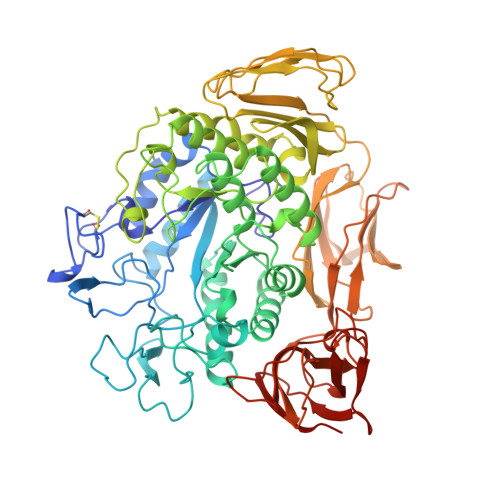
Substrate binding to a cyclodextrin glycosyltransferase and mutations increasing the gamma-cyclodextrin production.

Parsiegla, G.Schmidt, A.K.Schulz, G.E.

(1998) Eur J Biochem 255: 710-717

  • DOI: https://doi.org/10.1046/j.1432-1327.1998.2550710.x
  • Primary Citation Related Structures: 
    4CGT, 5CGT, 6CGT, 7CGT, 8CGT, 9CGT

  • PubMed Abstract: 

    Bacterial cyclodextrin glycosyltransferases use starch to produce cyclic maltooligosaccharides (cyclodextrins) which are of interest in various applications. The cyclization reaction gives rise to a spectrum of ring sizes consisting of predominantly six to eight glucosyl units. Using the enzyme from Bacillus circulans strain no. 8, binding studies have been performed with several substrates and analogues. The observed binding modes differ in detail, but agree in general with data on homologous enzymes. Based on these binding studies, two mutations were designed that changed the production spectrum from the predominant product beta-cyclodextrin of the wild-type enzyme towards gamma-cyclodextrin, which is of practical interest because it is rare and can encapsulate larger nonpolar compounds.


  • Organizational Affiliation
    • Institut für Organische Chemie und Biochemie, Freiburg im Breisgau, Germany.

Macromolecule Content 

  • Total Structure Weight: 75.47 kDa 
  • Atom Count: 5,516 
  • Modeled Residue Count: 684 
  • Deposited Residue Count: 684 
  • Unique protein chains: 1

Macromolecules

Find similar proteins by:|  3D Structure
Entity ID: 1
MoleculeChains  Sequence LengthOrganismDetailsImage
CYCLODEXTRIN GLYCOSYLTRANSFERASE684Niallia circulansMutation(s): 1 
EC: 2.4.1.19
UniProt
Find proteins for P30920 (Niallia circulans)
Explore P30920 
Go to UniProtKB:  P30920
Entity Groups
Sequence Clusters30% Identity50% Identity70% Identity90% Identity95% Identity100% Identity
UniProt GroupP30920
Sequence Annotations
Expand
Reference Sequence

Oligosaccharides

Help  
Entity ID: 2
MoleculeChains Length2D Diagram GlycosylationD Interactions
alpha-D-glucopyranose-(1-4)-beta-D-glucopyranose
B, C
2N/A
Glycosylation Resources
GlyTouCan: G66120GK
GlyCosmos: G66120GK

Biologically Interesting Molecules (External Reference) 

1 Unique

Experimental Data & Validation

Experimental Data

  • Method: X-RAY DIFFRACTION
  • Resolution: 2.50 Å
  • R-Value Free:  0.247 (Depositor) 
  • R-Value Work:  0.177 (Depositor), 0.184 (DCC) 
  • R-Value Observed: 0.177 (Depositor) 
Space Group: P 21 21 21
Unit Cell:
Length ( Å )Angle ( ˚ )
a = 93.5α = 90
b = 104.9β = 90
c = 113.8γ = 90
Software Package:
Software NamePurpose
XDSdata scaling
XSCALEdata scaling
X-PLORmodel building
X-PLORrefinement
XDSdata reduction
X-PLORphasing

Structure Validation

View Full Validation Report


Ligand Structure Quality Assessment 


Entry History 

Deposition Data

Revision History  (Full details and data files)

  • Version 1.0: 1998-08-12
    Type: Initial release
  • Version 1.1: 2008-03-25
    Changes: Version format compliance
  • Version 1.2: 2011-07-13
    Changes: Version format compliance
  • Version 2.0: 2020-07-29
    Type: Remediation
    Reason: Carbohydrate remediation
    Changes: Atomic model, Data collection, Derived calculations, Structure summary
  • Version 2.1: 2021-11-03
    Changes: Database references, Structure summary
  • Version 2.2: 2024-10-30
    Changes: Data collection, Structure summary