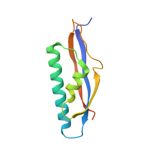
5CES | pdb_00005ces

C-terminal domain of the R-type pyocin baseplate protein PA0618


Experimental Data Snapshot

  • Method: X-RAY DIFFRACTION
  • Resolution: 2.10 Å
  • R-Value Free: 
    0.236 (Depositor), 0.248 (DCC) 
  • R-Value Work: 
    0.190 (Depositor), 0.201 (DCC) 
  • R-Value Observed: 
    0.192 (Depositor) 

wwPDB Validation 3D Report Full Report

Validation slider image for 5CES

This is version 1.5 of the entry. See complete history. 

Literature

Action of a minimal contractile bactericidal nanomachine.

Ge, P., Scholl, D., Prokhorov, N.S., Avaylon, J., Shneider, M.M., Browning, C., Buth, S.A., Plattner, M., Chakraborty, U., Ding, K., Leiman, P.G., Miller, J.F., Zhou, Z.H.

(2020) Nature 580: 658-662

  • DOI: https://doi.org/10.1038/s41586-020-2186-z
  • Primary Citation Related Structures: 
    5CES, 6PYT, 6U5B, 6U5F, 6U5H, 6U5J, 6U5K

  • PubMed Abstract: 

    R-type bacteriocins are minimal contractile nanomachines that hold promise as precision antibiotics 1-4 . Each bactericidal complex uses a collar to bridge a hollow tube with a contractile sheath loaded in a metastable state by a baseplate scaffold 1,2 . Fine-tuning of such nucleic acid-free protein machines for precision medicine calls for an atomic description of the entire complex and contraction mechanism, which is not available from baseplate structures of the (DNA-containing) T4 bacteriophage 5 . Here we report the atomic model of the complete R2 pyocin in its pre-contraction and post-contraction states, each containing 384 subunits of 11 unique atomic models of 10 gene products. Comparison of these structures suggests the following sequence of events during pyocin contraction: tail fibres trigger lateral dissociation of baseplate triplexes; the dissociation then initiates a cascade of events leading to sheath contraction; and this contraction converts chemical energy into mechanical force to drive the iron-tipped tube across the bacterial cell surface, killing the bacterium.


  • Organizational Affiliation: 
    • Department of Microbiology, Immunology and Molecular Genetics, University of California, Los Angeles (UCLA), Los Angeles, CA, USA.

Macromolecule Content 

  • Total Structure Weight: 22.6 kDa 
  • Atom Count: 1,461 
  • Modeled Residue Count: 182 
  • Deposited Residue Count: 204 
  • Unique protein chains: 1

Macromolecules

Find similar proteins by:|  3D Structure
Entity ID: 1
MoleculeChains  Sequence LengthOrganismDetailsImage
PA0618
A, B
102Pseudomonas aeruginosa PAO1Mutation(s): 0 
Gene Names: PA0618
UniProt
Find proteins for G3XCX5 (Pseudomonas aeruginosa (strain ATCC 15692 / DSM 22644 / CIP 104116 / JCM 14847 / LMG 12228 / 1C / PRS 101 / PAO1))
Explore G3XCX5 
Go to UniProtKB:  G3XCX5
Entity Groups
Sequence Clusters30% Identity50% Identity70% Identity90% Identity95% Identity100% Identity
UniProt GroupG3XCX5
Sequence Annotations
Expand
Reference Sequence

Experimental Data & Validation

Experimental Data

  • Method: X-RAY DIFFRACTION
  • Resolution: 2.10 Å
  • R-Value Free:  0.236 (Depositor), 0.248 (DCC) 
  • R-Value Work:  0.190 (Depositor), 0.201 (DCC) 
  • R-Value Observed: 0.192 (Depositor) 
Space Group: P 43
Unit Cell:
Length ( Å )Angle ( ˚ )
a = 72.611α = 90
b = 72.611β = 90
c = 46.519γ = 90
Software Package:
Software NamePurpose
PHENIXrefinement
XDSdata reduction
XDSdata scaling
MOLREPphasing

Structure Validation

View Full Validation Report


Entry History 

& Funding Information

Deposition Data


Funding OrganizationLocationGrant Number
Swiss National Science FoundationSwitzerland310030_144243

Revision History  (Full details and data files)

  • Version 1.0: 2016-07-27
    Type: Initial release
  • Version 1.1: 2018-05-16
    Changes: Data collection, Structure summary
  • Version 1.2: 2018-05-23
    Changes: Data collection, Database references, Structure summary
  • Version 1.3: 2020-04-29
    Changes: Database references
  • Version 1.4: 2020-05-13
    Changes: Database references
  • Version 1.5: 2024-05-08
    Changes: Data collection, Database references