5A9X | pdb_00005a9x

Structure of GDP bound BipA


Experimental Data Snapshot

  • Method: X-RAY DIFFRACTION
  • Resolution: 3.80 Å
  • R-Value Free: 
    0.313 (Depositor), 0.321 (DCC) 
  • R-Value Work: 
    0.248 (Depositor), 0.268 (DCC) 
  • R-Value Observed: 
    0.254 (Depositor) 

wwPDB Validation 3D Report Full Report

Validation slider image for 5A9X

Ligand Structure Quality Assessment 


This is version 1.3 of the entry. See complete history

Literature

Structure of Bipa in GTP Form Bound to the Ratcheted Ribosome.

Kumar, V.Chen, Y.Ero, R.Ahmed, T.Tan, J.Li, Z.Wong, A.S.W.Bhushan, S.Gao, Y.

(2015) Proc Natl Acad Sci U S A 112: 10944

  • DOI: https://doi.org/10.1073/pnas.1513216112
  • Primary Citation Related Structures: 
    5A9V, 5A9W, 5A9X, 5A9Y, 5A9Z, 5AA0

  • PubMed Abstract: 

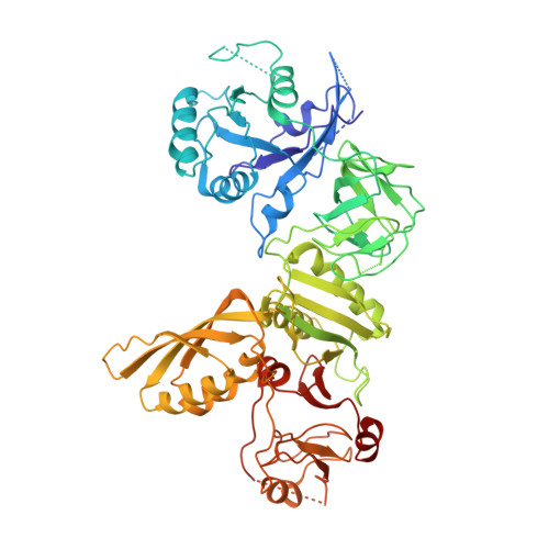
    BPI-inducible protein A (BipA) is a member of the family of ribosome-dependent translational GTPase (trGTPase) factors along with elongation factors G and 4 (EF-G and EF4). Despite being highly conserved in bacteria and playing a critical role in coordinating cellular responses to environmental changes, its structures (isolated and ribosome bound) remain elusive. Here, we present the crystal structures of apo form and GTP analog, GDP, and guanosine-3',5'-bisdiphosphate (ppGpp)-bound BipA. In addition to having a distinctive domain arrangement, the C-terminal domain of BipA has a unique fold. Furthermore, we report the cryo-electron microscopy structure of BipA bound to the ribosome in its active GTP form and elucidate the unique structural attributes of BipA interactions with the ribosome and A-site tRNA in the light of its possible function in regulating translation.


  • Organizational Affiliation
    • Institute of Molecular and Cell Biology, A*STAR, 138673, Singapore; School of Biological Sciences, Nanyang Technological University, 637551, Singapore.

Macromolecule Content 

  • Total Structure Weight: 67.88 kDa 
  • Atom Count: 4,167 
  • Modeled Residue Count: 556 
  • Deposited Residue Count: 607 
  • Unique protein chains: 1

Macromolecules

Find similar proteins by:|  3D Structure
Entity ID: 1
MoleculeChains  Sequence LengthOrganismDetailsImage
GTP-BINDING PROTEIN607Escherichia coliMutation(s): 0 
EC: 3.6.5
UniProt
Find proteins for P0A3B2 (Escherichia coli O127:H6 (strain E2348/69 / EPEC))
Explore P0A3B2 
Go to UniProtKB:  P0A3B2
Entity Groups
Sequence Clusters30% Identity50% Identity70% Identity90% Identity95% Identity100% Identity
UniProt GroupP0A3B2
Sequence Annotations
Expand
Reference Sequence

Small Molecules

Ligands 1 Unique
IDChains Name / Formula / InChI Key2D Diagram3D Interactions
GDP

Query on GDP


Download:Ideal Coordinates CCD File
B [auth A]GUANOSINE-5'-DIPHOSPHATE
C10 H15 N5 O11 P2
QGWNDRXFNXRZMB-UUOKFMHZSA-N

Experimental Data & Validation

Experimental Data

  • Method: X-RAY DIFFRACTION
  • Resolution: 3.80 Å
  • R-Value Free:  0.313 (Depositor), 0.321 (DCC) 
  • R-Value Work:  0.248 (Depositor), 0.268 (DCC) 
  • R-Value Observed: 0.254 (Depositor) 
Space Group: I 41 3 2
Unit Cell:
Length ( Å )Angle ( ˚ )
a = 241.946α = 90
b = 241.946β = 90
c = 241.946γ = 90
Software Package:
Software NamePurpose
REFMACrefinement
XDSdata reduction
XDSdata scaling

Structure Validation

View Full Validation Report


Ligand Structure Quality Assessment 


Entry History 

Deposition Data

Revision History  (Full details and data files)

  • Version 1.0: 2015-08-26
    Type: Initial release
  • Version 1.1: 2015-09-02
    Changes: Database references
  • Version 1.2: 2015-09-16
    Changes: Database references
  • Version 1.3: 2024-05-08
    Changes: Data collection, Database references, Derived calculations, Other