5A2Y | pdb_00005a2y

Crystal structure of mtPAP in complex with UTP


Experimental Data Snapshot

  • Method: X-RAY DIFFRACTION
  • Resolution: 2.75 Å
  • R-Value Free: 
    0.255 (Depositor), 0.247 (DCC) 
  • R-Value Work: 
    0.216 (Depositor), 0.214 (DCC) 
  • R-Value Observed: 
    0.218 (Depositor) 

wwPDB Validation 3D Report Full Report

Validation slider image for 5A2Y

Ligand Structure Quality Assessment 


This is version 1.3 of the entry. See complete history

Literature

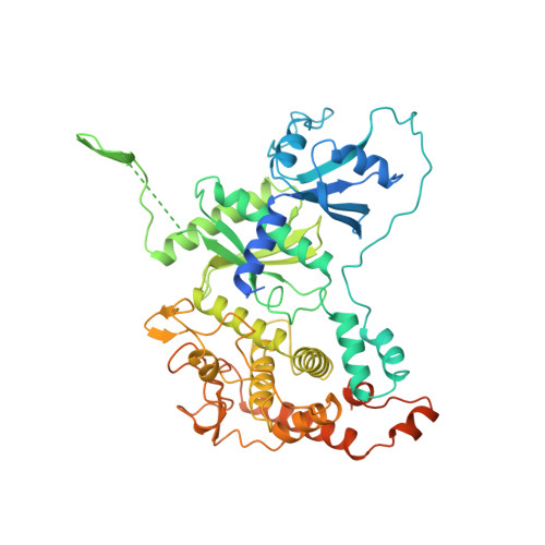
Structure of Mitochondrial Poly(A) RNA Polymerase Reveals the Structural Basis for Dimerization, ATP Selectivity and the Spax4 Disease Phenotype.

Lapkouski, M.Hallberg, B.M.

(2015) Nucleic Acids Res 43: 9065

  • DOI: https://doi.org/10.1093/nar/gkv861
  • Primary Citation Related Structures: 
    5A2V, 5A2W, 5A2X, 5A2Y, 5A2Z, 5A30

  • PubMed Abstract: 

    Polyadenylation, performed by poly(A) polymerases (PAPs), is a ubiquitous post-transcriptional modification that plays key roles in multiple aspects of RNA metabolism. Although cytoplasmic and nuclear PAPs have been studied extensively, the mechanism by which mitochondrial PAP (mtPAP) selects adenosine triphosphate over other nucleotides is unknown. Furthermore, mtPAP is unique because it acts as a dimer. However, mtPAP's dimerization requirement remains enigmatic. Here, we show the structural basis for mtPAP's nucleotide selectivity, dimerization and catalysis. Our structures reveal an intricate dimerization interface that features an RNA-recognition module formed through strand complementation. Further, we propose the structural basis for the N478D mutation that drastically reduces the length of poly(A) tails on mitochondrial mRNAs in patients with spastic ataxia 4 (SPAX4), a severe and progressive neurodegenerative disease.


  • Organizational Affiliation
    • Department of Cell and Molecular Biology, Karolinska Institutet, 171 77 Stockholm, Sweden Röntgen-Ångström-Cluster, Karolinska Institutet Outstation, Centre for Structural Systems Biology, DESY-Campus, 22607 Hamburg, Germany.

Macromolecule Content 

  • Total Structure Weight: 126.93 kDa 
  • Atom Count: 7,501 
  • Modeled Residue Count: 932 
  • Deposited Residue Count: 1,110 
  • Unique protein chains: 1

Macromolecules

Find similar proteins by:|  3D Structure
Entity ID: 1
MoleculeChains  Sequence LengthOrganismDetailsImage
MITOCHONDRIAL PROTEIN
A, B
555Gallus gallusMutation(s): 0 
UniProt
Find proteins for F1NBW0 (Gallus gallus)
Explore F1NBW0 
Go to UniProtKB:  F1NBW0
Entity Groups
Sequence Clusters30% Identity50% Identity70% Identity90% Identity95% Identity100% Identity
UniProt GroupF1NBW0
Sequence Annotations
Expand
Reference Sequence

Experimental Data & Validation

Experimental Data

  • Method: X-RAY DIFFRACTION
  • Resolution: 2.75 Å
  • R-Value Free:  0.255 (Depositor), 0.247 (DCC) 
  • R-Value Work:  0.216 (Depositor), 0.214 (DCC) 
  • R-Value Observed: 0.218 (Depositor) 
Space Group: P 21 21 21
Unit Cell:
Length ( Å )Angle ( ˚ )
a = 59.71α = 90
b = 93.83β = 90
c = 192.436γ = 90
Software Package:
Software NamePurpose
REFMACrefinement
SCALEPACKdata scaling
MOLREPphasing

Structure Validation

View Full Validation Report


Ligand Structure Quality Assessment 


Entry History 

Deposition Data

Revision History  (Full details and data files)

  • Version 1.0: 2015-09-09
    Type: Initial release
  • Version 1.1: 2015-10-28
    Changes: Database references
  • Version 1.2: 2017-08-23
    Changes: Data collection
  • Version 1.3: 2024-05-08
    Changes: Data collection, Database references, Derived calculations, Other