4CIS

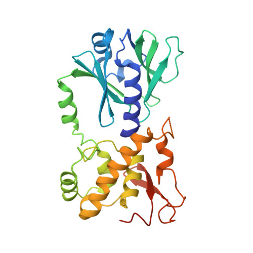
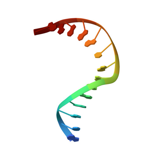
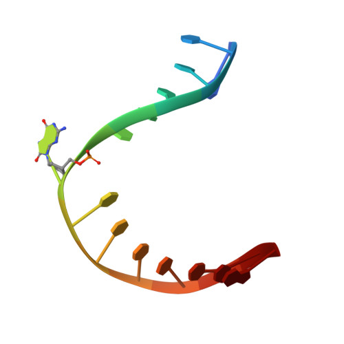
Structure of MutM in complex with carbocyclic 8-oxo-G containing DNA


Experimental Data Snapshot

  • Method: X-RAY DIFFRACTION
  • Resolution: 2.05 Å
  • R-Value Free: 0.239 
  • R-Value Work: 0.214 
  • R-Value Observed: 0.215 

Starting Model: experimental
View more details

wwPDB Validation   3D Report Full Report


This is version 2.1 of the entry. See complete history


Literature

Ribose-protonated DNA base excision repair: a combined theoretical and experimental study.

Sadeghian, K.Flaig, D.Blank, I.D.Schneider, S.Strasser, R.Stathis, D.Winnacker, M.Carell, T.Ochsenfeld, C.

(2014) Angew Chem Int Ed Engl 53: 10044-10048

  • DOI: https://doi.org/10.1002/anie.201403334
  • Primary Citation of Related Structures:  
    4CIS

  • PubMed Abstract: 

    Living organisms protect the genome against external influences by recognizing and repairing damaged DNA. A common source of gene mutation is the oxidized guanine, which undergoes base excision repair through cleavage of the glycosidic bond between the ribose and the nucleobase of the lesion. We unravel the repair mechanism utilized by bacterial glycosylase, MutM, using quantum-chemical calculations involving more than 1000 atoms of the catalytic site. In contrast to the base-protonated pathway currently favored in the literature, we show that the initial protonation of the lesion's ribose paves the way for an almost barrier-free glycosidic cleavage. The combination of theoretical and experimental data provides further insight into the selectivity and discrimination of MutM's binding site toward various substrates.


  • Organizational Affiliation

    Chair of Theoretical Chemistry, Department of Chemistry, University of Munich (LMU), Butenandtstrasse 7, 81377 Munich (Germany); Center for Integrated Protein Science (CIPSM) at the Department of Chemistry, University of Munich (LMU), Butenandtstrasse 5-13, 81377 Munich (Germany).


Macromolecules

Find similar proteins by:  (by identity cutoff)  |  3D Structure
Entity ID: 1
MoleculeChains Sequence LengthOrganismDetailsImage
FORMAMIDOPYRIMIDIN DNA GLYCOSYLASE
A, B
283Lactococcus cremorisMutation(s): 0 
EC: 3.2.2.23 (PDB Primary Data), 4.2.99.18 (UniProt)
UniProt
Find proteins for Q031W6 (Lactococcus lactis subsp. cremoris (strain SK11))
Explore Q031W6 
Go to UniProtKB:  Q031W6
Entity Groups  
Sequence Clusters30% Identity50% Identity70% Identity90% Identity95% Identity100% Identity
UniProt GroupQ031W6
Sequence Annotations
Expand
  • Reference Sequence

Find similar nucleic acids by:  Sequence   |   3D Structure  

Entity ID: 2
MoleculeChains LengthOrganismImage
DNA14synthetic construct
Sequence Annotations
Expand
  • Reference Sequence

Find similar nucleic acids by:  Sequence   |   3D Structure  

Entity ID: 3
MoleculeChains LengthOrganismImage
DNA14synthetic construct
Sequence Annotations
Expand
  • Reference Sequence
Experimental Data & Validation

Experimental Data

  • Method: X-RAY DIFFRACTION
  • Resolution: 2.05 Å
  • R-Value Free: 0.239 
  • R-Value Work: 0.214 
  • R-Value Observed: 0.215 
  • Space Group: P 21 21 21
Unit Cell:
Length ( Å )Angle ( ˚ )
a = 43.063α = 90
b = 112.692β = 90
c = 132.882γ = 90
Software Package:
Software NamePurpose
REFMACrefinement
XDSdata reduction
XSCALEdata scaling
PHASERphasing

Structure Validation

View Full Validation Report


Entry History 

Deposition Data

Revision History  (Full details and data files)

  • Version 1.0: 2014-06-04
    Type: Initial release
  • Version 1.1: 2014-08-06
    Changes: Database references
  • Version 1.2: 2014-09-17
    Changes: Database references
  • Version 2.0: 2018-03-07
    Changes: Atomic model, Database references, Source and taxonomy
  • Version 2.1: 2023-12-20
    Changes: Data collection, Database references, Derived calculations, Other, Refinement description