4YRJ | pdb_00004yrj

Crystal structure of T. cruzi Histidyl-tRNA synthetase in complex with 4-chlorobenzene-1,2-diamine (Chem 256)


Experimental Data Snapshot

  • Method: X-RAY DIFFRACTION
  • Resolution: 2.30 Å
  • R-Value Free: 
    0.245 (Depositor), 0.245 (DCC) 
  • R-Value Work: 
    0.205 (Depositor), 0.206 (DCC) 
  • R-Value Observed: 
    0.207 (Depositor) 

Starting Model: experimental
View more details

wwPDB Validation 3D Report Full Report

Validation slider image for 4YRJ

Ligand Structure Quality Assessment 


This is version 1.3 of the entry. See complete history

Literature

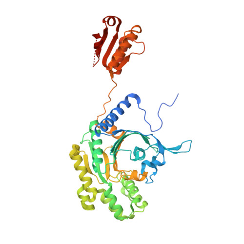
A binding hotspot in Trypanosoma cruzi histidyl-tRNA synthetase revealed by fragment-based crystallographic cocktail screens.

Koh, C.Y.Siddaramaiah, L.K.Ranade, R.M.Nguyen, J.Jian, T.Zhang, Z.Gillespie, J.R.Buckner, F.S.Verlinde, C.L.Fan, E.Hol, W.G.

(2015) Acta Crystallogr D Biol Crystallogr 71: 1684-1698

  • DOI: https://doi.org/10.1107/S1399004715007683
  • Primary Citation Related Structures: 
    4YP0, 4YPF, 4YRC, 4YRE, 4YRF, 4YRG, 4YRI, 4YRJ, 4YRK, 4YRL, 4YRM, 4YRN, 4YRO, 4YRP, 4YRQ, 4YRR, 4YRS, 4YRT

  • PubMed Abstract: 

    American trypanosomiasis, commonly known as Chagas disease, is a neglected tropical disease caused by the protozoan parasite Trypanosoma cruzi. The chronic form of the infection often causes debilitating morbidity and mortality. However, the current treatment for the disease is typically inadequate owing to drug toxicity and poor efficacy, necessitating a continual effort to discover and develop new antiparasitic therapeutic agents. The structure of T. cruzi histidyl-tRNA synthetase (HisRS), a validated drug target, has previously been reported. Based on this structure and those of human cytosolic HisRS, opportunities for the development of specific inhibitors were identified. Here, efforts are reported to identify small molecules that bind to T. cruzi HisRS through fragment-based crystallographic screening in order to arrive at chemical starting points for the development of specific inhibitors. T. cruzi HisRS was soaked into 68 different cocktails from the Medical Structural Genomics of Pathogenic Protozoa (MSGPP) fragment library and diffraction data were collected to identify bound fragments after soaking. A total of 15 fragments were identified, all bound to the same site on the protein, revealing a fragment-binding hotspot adjacent to the ATP-binding pocket. On the basis of the initial hits, the design of reactive fragments targeting the hotspot which would be simultaneously covalently linked to a cysteine residue present only in trypanosomatid HisRS was initiated. Inhibition of T. cruzi HisRS was observed with the resultant reactive fragments and the anticipated binding mode was confirmed crystallographically. These results form a platform for the development of future generations of selective inhibitors for trypanosomatid HisRS.


  • Organizational Affiliation
    • Department of Biochemistry, University of Washington, Seattle, WA 98195, USA.

Macromolecule Content 

  • Total Structure Weight: 51.99 kDa 
  • Atom Count: 3,345 
  • Modeled Residue Count: 413 
  • Deposited Residue Count: 456 
  • Unique protein chains: 1

Macromolecules

Find similar proteins by:|  3D Structure
Entity ID: 1
MoleculeChains  Sequence LengthOrganismDetailsImage
Histidyl-tRNA synthetase456Trypanosoma cruzi strain CL BrenerMutation(s): 0 
Gene Names: Tc00.1047053507019.40
EC: 6.1.1.21
UniProt
Find proteins for Q4DA54 (Trypanosoma cruzi (strain CL Brener))
Explore Q4DA54 
Go to UniProtKB:  Q4DA54
Entity Groups
Sequence Clusters30% Identity50% Identity70% Identity90% Identity95% Identity100% Identity
UniProt GroupQ4DA54
Sequence Annotations
Expand
Reference Sequence

Small Molecules

Ligands 5 Unique
IDChains Name / Formula / InChI Key2D Diagram3D Interactions
HIS

Query on HIS


Download:Ideal Coordinates CCD File
B [auth A]HISTIDINE
C6 H10 N3 O2
HNDVDQJCIGZPNO-YFKPBYRVSA-O
4JJ

Query on 4JJ


Download:Ideal Coordinates CCD File
C [auth A]4-chlorobenzene-1,2-diamine
C6 H7 Cl N2
BXIXXXYDDJVHDL-UHFFFAOYSA-N
SO4

Query on SO4


Download:Ideal Coordinates CCD File
G [auth A],
H [auth A],
I [auth A]
SULFATE ION
O4 S
QAOWNCQODCNURD-UHFFFAOYSA-L
DMS

Query on DMS


Download:Ideal Coordinates CCD File
D [auth A],
E [auth A]
DIMETHYL SULFOXIDE
C2 H6 O S
IAZDPXIOMUYVGZ-UHFFFAOYSA-N
EDO

Query on EDO


Download:Ideal Coordinates CCD File
F [auth A]1,2-ETHANEDIOL
C2 H6 O2
LYCAIKOWRPUZTN-UHFFFAOYSA-N

Experimental Data & Validation

Experimental Data

  • Method: X-RAY DIFFRACTION
  • Resolution: 2.30 Å
  • R-Value Free:  0.245 (Depositor), 0.245 (DCC) 
  • R-Value Work:  0.205 (Depositor), 0.206 (DCC) 
  • R-Value Observed: 0.207 (Depositor) 
Space Group: I 1 2 1
Unit Cell:
Length ( Å )Angle ( ˚ )
a = 64.875α = 90
b = 119.306β = 92.61
c = 66.23γ = 90
Software Package:
Software NamePurpose
DENZOdata reduction
Aimlessdata scaling
REFMACrefinement
PDB_EXTRACTdata extraction
HKL-2000data reduction
REFMACphasing

Structure Validation

View Full Validation Report


Ligand Structure Quality Assessment 


Entry History 

Deposition Data

Revision History  (Full details and data files)

  • Version 1.0: 2015-08-12
    Type: Initial release
  • Version 1.1: 2017-11-22
    Changes: Derived calculations, Refinement description
  • Version 1.2: 2018-04-18
    Changes: Data collection, Database references, Derived calculations, Structure summary
  • Version 1.3: 2023-09-27
    Changes: Data collection, Database references, Derived calculations, Refinement description, Structure summary