4YN2 | pdb_00004yn2

THE ATOMIC STRUCTURE OF WISEANA SPP ENTOMOPOXVIRUS (WSEPV) FUSOLIN SPINDLES


Experimental Data Snapshot

  • Method: X-RAY DIFFRACTION
  • Resolution: 2.02 Å
  • R-Value Free: 
    0.199 (Depositor), 0.200 (DCC) 
  • R-Value Work: 
    0.166 (Depositor) 
  • R-Value Observed: 
    0.168 (Depositor) 

Starting Model: other
View more details

wwPDB Validation 3D Report Full Report

Validation slider image for 4YN2

This is version 2.1 of the entry. See complete history

Literature

Structural basis for the enhancement of virulence by viral spindles and their in vivo crystallization.

Chiu, E.Hijnen, M.Bunker, R.D.Boudes, M.Rajendran, C.Aizel, K.Olieric, V.Schulze-Briese, C.Mitsuhashi, W.Young, V.Ward, V.K.Bergoin, M.Metcalf, P.Coulibaly, F.

(2015) Proc Natl Acad Sci U S A 112: 3973-3978

  • DOI: https://doi.org/10.1073/pnas.1418798112
  • Primary Citation Related Structures: 
    4OW5, 4X27, 4X29, 4YN1, 4YN2

  • PubMed Abstract: 

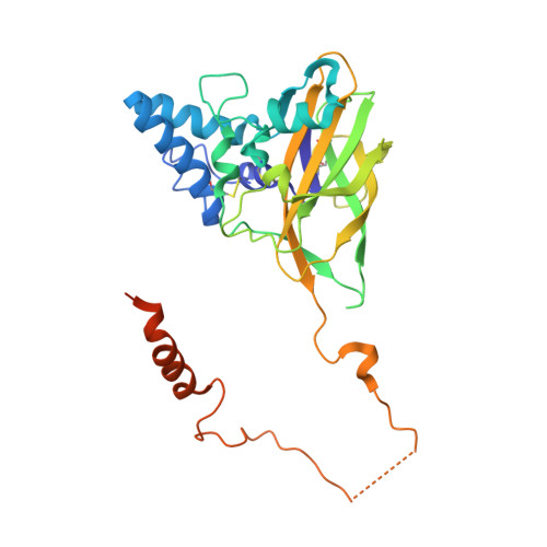
    The great benefits that chemical pesticides have brought to agriculture are partly offset by widespread environmental damage to nontarget species and threats to human health. Microbial bioinsecticides are considered safe and highly specific alternatives but generally lack potency. Spindles produced by insect poxviruses are crystals of the fusolin protein that considerably boost not only the virulence of these viruses but also, in cofeeding experiments, the insecticidal activity of unrelated pathogens. However, the mechanisms by which spindles assemble into ultra-stable crystals and enhance virulence are unknown. Here we describe the structure of viral spindles determined by X-ray microcrystallography from in vivo crystals purified from infected insects. We found that a C-terminal molecular arm of fusolin mediates the assembly of a globular domain, which has the hallmarks of lytic polysaccharide monooxygenases of chitinovorous bacteria. Explaining their unique stability, a 3D network of disulfide bonds between fusolin dimers covalently crosslinks the entire crystalline matrix of spindles. However, upon ingestion by a new host, removal of the molecular arm abolishes this stabilizing network leading to the dissolution of spindles. The released monooxygenase domain is then free to disrupt the chitin-rich peritrophic matrix that protects insects against oral infections. The mode of action revealed here may guide the design of potent spindles as synergetic additives to bioinsecticides.


  • Organizational Affiliation
    • School of Biological Sciences, University of Auckland, Auckland, 1010, New Zealand;

Macromolecule Content 

  • Total Structure Weight: 37 kDa 
  • Atom Count: 2,481 
  • Modeled Residue Count: 281 
  • Deposited Residue Count: 313 
  • Unique protein chains: 1

Macromolecules

Find similar proteins by:|  3D Structure
Entity ID: 1
MoleculeChains  Sequence LengthOrganismDetailsImage
FUSOLIN313unidentified entomopoxvirusMutation(s): 0 
UniProt
Find proteins for A0A0J9X287 (unidentified entomopoxvirus)
Explore A0A0J9X287 
Go to UniProtKB:  A0A0J9X287
Entity Groups
Sequence Clusters30% Identity50% Identity70% Identity90% Identity95% Identity100% Identity
UniProt GroupA0A0J9X287
Glycosylation
Glycosylation Sites: 1
Sequence Annotations
Expand
Reference Sequence

Oligosaccharides

Help  
Entity ID: 2
MoleculeChains Length2D Diagram GlycosylationD Interactions
beta-D-mannopyranose-(1-4)-2-acetamido-2-deoxy-beta-D-glucopyranose-(1-4)-2-acetamido-2-deoxy-beta-D-glucopyranose
B
3N-Glycosylation
Glycosylation Resources
GlyTouCan: G15407YE
GlyCosmos: G15407YE
GlyGen: G15407YE

Experimental Data & Validation

Experimental Data

  • Method: X-RAY DIFFRACTION
  • Resolution: 2.02 Å
  • R-Value Free:  0.199 (Depositor), 0.200 (DCC) 
  • R-Value Work:  0.166 (Depositor) 
  • R-Value Observed: 0.168 (Depositor) 
Space Group: P 41 21 2
Unit Cell:
Length ( Å )Angle ( ˚ )
a = 68.182α = 90
b = 68.182β = 90
c = 128.058γ = 90
Software Package:
Software NamePurpose
DENZOdata reduction
SCALEPACKdata scaling
PHASERphasing
SHELXDEphasing
ARPmodel building
Cootmodel building
BUSTERrefinement
SHARPphasing

Structure Validation

View Full Validation Report


Entry History 

Deposition Data

Revision History  (Full details and data files)

  • Version 1.0: 2015-04-08
    Type: Initial release
  • Version 1.1: 2015-04-15
    Changes: Database references
  • Version 1.2: 2017-11-22
    Changes: Database references, Derived calculations, Refinement description, Source and taxonomy
  • Version 2.0: 2020-07-29
    Type: Remediation
    Reason: Carbohydrate remediation
    Changes: Atomic model, Data collection, Derived calculations, Structure summary
  • Version 2.1: 2024-10-23
    Changes: Data collection, Database references, Refinement description, Structure summary