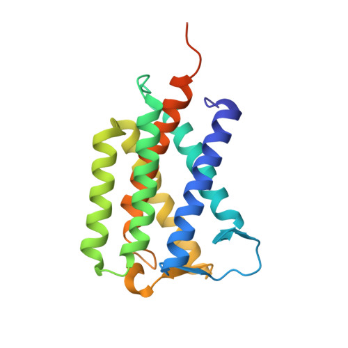
4XU6

Crystal structure of cross-linked MvINS R77C trimer at 1.9A resolution


Experimental Data Snapshot

  • Method: X-RAY DIFFRACTION
  • Resolution: 1.90 Å
  • R-Value Free: 0.203 
  • R-Value Work: 0.189 
  • R-Value Observed: 0.190 

wwPDB Validation   3D Report Full Report


Ligand Structure Quality Assessment 


This is version 1.2 of the entry. See complete history


Literature

PROTEIN STRUCTURE. Crystal structure of a mycobacterial Insig homolog provides insight into how these sensors monitor sterol levels

Ren, R.Zhou, X.He, Y.Ke, M.Wu, J.Liu, X.Yan, C.Wu, Y.Gong, X.Lei, X.Yan, S.F.Radhakrishnan, A.Yan, N.

(2015) Science 349: 187-191

  • DOI: https://doi.org/10.1126/science.aab1091
  • Primary Citation of Related Structures:  
    4XU4, 4XU5, 4XU6

  • PubMed Abstract: 

    Insulin-induced gene 1 (Insig-1) and Insig-2 are endoplasmic reticulum membrane-embedded sterol sensors that regulate the cellular accumulation of sterols. Despite their physiological importance, the structural information on Insigs remains limited. Here we report the high-resolution structures of MvINS, an Insig homolog from Mycobacterium vanbaalenii. MvINS exists as a homotrimer. Each protomer comprises six transmembrane segments (TMs), with TM3 and TM4 contributing to homotrimerization. The six TMs enclose a V-shaped cavity that can accommodate a diacylglycerol molecule. A homology-based structural model of human Insig-2, together with biochemical characterizations, suggest that the central cavity of Insig-2 accommodates 25-hydroxycholesterol, whereas TM3 and TM4 engage in Scap binding. These analyses provide an important framework for further functional and mechanistic understanding of Insig proteins and the sterol regulatory element-binding protein pathway.


  • Organizational Affiliation

    State Key Laboratory of Membrane Biology, Tsinghua University, Beijing 100084, China. Center for Structural Biology, School of Life Sciences, School of Medicine, Tsinghua University, Beijing 100084, China. Tsinghua-Peking Center for Life Sciences, Tsinghua University, Beijing 100084, China.


Macromolecules
Find similar proteins by:  (by identity cutoff)  |  3D Structure
Entity ID: 1
MoleculeChains Sequence LengthOrganismDetailsImage
Uncharacterized protein210Mycolicibacterium vanbaalenii PYR-1Mutation(s): 0 
Gene Names: Mvan_1475
Membrane Entity: Yes 
UniProt
Find proteins for A1T557 (Mycolicibacterium vanbaalenii (strain DSM 7251 / JCM 13017 / BCRC 16820 / KCTC 9966 / NRRL B-24157 / PYR-1))
Explore A1T557 
Go to UniProtKB:  A1T557
Entity Groups  
Sequence Clusters30% Identity50% Identity70% Identity90% Identity95% Identity100% Identity
UniProt GroupA1T557
Sequence Annotations
Expand
  • Reference Sequence
Experimental Data & Validation

Experimental Data

  • Method: X-RAY DIFFRACTION
  • Resolution: 1.90 Å
  • R-Value Free: 0.203 
  • R-Value Work: 0.189 
  • R-Value Observed: 0.190 
  • Space Group: H 3
Unit Cell:
Length ( Å )Angle ( ˚ )
a = 85.596α = 90
b = 85.596β = 90
c = 126.12γ = 120
Software Package:
Software NamePurpose
PHENIXrefinement
HKL-2000data reduction
HKL-2000data scaling
PHASERphasing

Structure Validation

View Full Validation Report


Ligand Structure Quality Assessment 


Entry History 

Deposition Data

Revision History  (Full details and data files)

  • Version 1.0: 2015-10-14
    Type: Initial release
  • Version 1.1: 2020-07-29
    Type: Remediation
    Reason: Carbohydrate remediation
    Changes: Data collection, Derived calculations, Structure summary
  • Version 1.2: 2024-03-20
    Changes: Data collection, Database references, Structure summary