4XU5 | pdb_00004xu5

Crystal structure of MvINS bound to a bromine-derived 14C Diacylglycerol (DAG) at 2.1A resolution


Experimental Data Snapshot

  • Method: X-RAY DIFFRACTION
  • Resolution: 2.10 Å
  • R-Value Free: 
    0.225 (Depositor), 0.226 (DCC) 
  • R-Value Work: 
    0.200 (Depositor) 
  • R-Value Observed: 
    0.201 (Depositor) 

wwPDB Validation 3D Report Full Report

Validation slider image for 4XU5

Ligand Structure Quality Assessment 


This is version 1.2 of the entry. See complete history

Literature

PROTEIN STRUCTURE. Crystal structure of a mycobacterial Insig homolog provides insight into how these sensors monitor sterol levels

Ren, R.Zhou, X.He, Y.Ke, M.Wu, J.Liu, X.Yan, C.Wu, Y.Gong, X.Lei, X.Yan, S.F.Radhakrishnan, A.Yan, N.

(2015) Science 349: 187-191

  • DOI: https://doi.org/10.1126/science.aab1091
  • Primary Citation Related Structures: 
    4XU4, 4XU5, 4XU6

  • PubMed Abstract: 

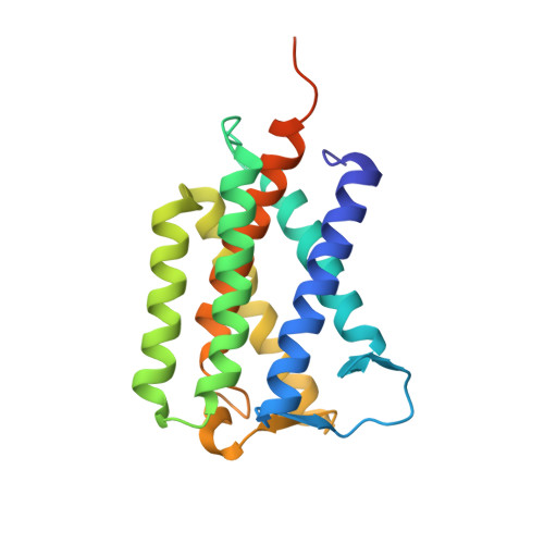
    Insulin-induced gene 1 (Insig-1) and Insig-2 are endoplasmic reticulum membrane-embedded sterol sensors that regulate the cellular accumulation of sterols. Despite their physiological importance, the structural information on Insigs remains limited. Here we report the high-resolution structures of MvINS, an Insig homolog from Mycobacterium vanbaalenii. MvINS exists as a homotrimer. Each protomer comprises six transmembrane segments (TMs), with TM3 and TM4 contributing to homotrimerization. The six TMs enclose a V-shaped cavity that can accommodate a diacylglycerol molecule. A homology-based structural model of human Insig-2, together with biochemical characterizations, suggest that the central cavity of Insig-2 accommodates 25-hydroxycholesterol, whereas TM3 and TM4 engage in Scap binding. These analyses provide an important framework for further functional and mechanistic understanding of Insig proteins and the sterol regulatory element-binding protein pathway.


  • Organizational Affiliation
    • State Key Laboratory of Membrane Biology, Tsinghua University, Beijing 100084, China. Center for Structural Biology, School of Life Sciences, School of Medicine, Tsinghua University, Beijing 100084, China. Tsinghua-Peking Center for Life Sciences, Tsinghua University, Beijing 100084, China.

Macromolecule Content 

  • Total Structure Weight: 23.13 kDa 
  • Atom Count: 1,516 
  • Modeled Residue Count: 189 
  • Deposited Residue Count: 210 
  • Unique protein chains: 1

Macromolecules

Find similar proteins by:|  3D Structure
Entity ID: 1
MoleculeChains  Sequence LengthOrganismDetailsImage
Uncharacterized protein210Mycolicibacterium vanbaalenii PYR-1Mutation(s): 0 
Gene Names: Mvan_1475
Membrane Entity: Yes 
UniProt
Find proteins for A1T557 (Mycolicibacterium vanbaalenii (strain DSM 7251 / JCM 13017 / BCRC 16820 / KCTC 9966 / NRRL B-24157 / PYR-1))
Explore A1T557 
Go to UniProtKB:  A1T557
Entity Groups
Sequence Clusters30% Identity50% Identity70% Identity90% Identity95% Identity100% Identity
UniProt GroupA1T557
Sequence Annotations
Expand
Reference Sequence

Small Molecules

Ligands 3 Unique
IDChains Name / Formula / InChI Key2D Diagram3D Interactions
LBR

Query on LBR


Download:Ideal Coordinates CCD File
B [auth A](2S)-1-[(13-bromotridecanoyl)oxy]-3-hydroxypropan-2-yl tetradecanoate
C30 H57 Br O5
VHCCBJMTBOAHLF-NDEPHWFRSA-N
BOG

Query on BOG


Download:Ideal Coordinates CCD File
H [auth A]octyl beta-D-glucopyranoside
C14 H28 O6
HEGSGKPQLMEBJL-RKQHYHRCSA-N
D10

Query on D10


Download:Ideal Coordinates CCD File
C [auth A],
D [auth A],
E [auth A],
F [auth A],
G [auth A]
DECANE
C10 H22
DIOQZVSQGTUSAI-UHFFFAOYSA-N

Experimental Data & Validation

Experimental Data

  • Method: X-RAY DIFFRACTION
  • Resolution: 2.10 Å
  • R-Value Free:  0.225 (Depositor), 0.226 (DCC) 
  • R-Value Work:  0.200 (Depositor) 
  • R-Value Observed: 0.201 (Depositor) 
Space Group: H 3
Unit Cell:
Length ( Å )Angle ( ˚ )
a = 83.393α = 90
b = 83.393β = 90
c = 124.885γ = 120
Software Package:
Software NamePurpose
PHENIXrefinement
HKL-2000data reduction
HKL-2000data scaling
PHASERphasing

Structure Validation

View Full Validation Report


Ligand Structure Quality Assessment 


Entry History 

Deposition Data

Revision History  (Full details and data files)

  • Version 1.0: 2015-10-14
    Type: Initial release
  • Version 1.1: 2020-07-29
    Type: Remediation
    Reason: Carbohydrate remediation
    Changes: Data collection, Derived calculations, Structure summary
  • Version 1.2: 2024-03-20
    Changes: Data collection, Database references, Structure summary