4XTL | pdb_00004xtl

Crystal structure of the light-driven sodium pump KR2 in the monomeric blue form, pH 4.3


Experimental Data Snapshot

  • Method: X-RAY DIFFRACTION
  • Resolution: 1.45 Å
  • R-Value Free: 
    0.216 (Depositor), 0.222 (DCC) 
  • R-Value Work: 
    0.187 (Depositor), 0.194 (DCC) 
  • R-Value Observed: 
    0.189 (Depositor) 

Starting Model: experimental
View more details

wwPDB Validation 3D Report Full Report

Validation slider image for 4XTL

Ligand Structure Quality Assessment 


This is version 1.5 of the entry. See complete history

Literature

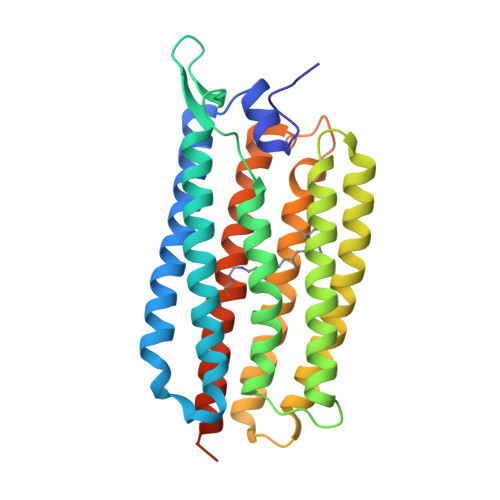
Crystal structure of a light-driven sodium pump.

Gushchin, I.Shevchenko, V.Polovinkin, V.Kovalev, K.Alekseev, A.Round, E.Borshchevskiy, V.Balandin, T.Popov, A.Gensch, T.Fahlke, C.Bamann, C.Willbold, D.Buldt, G.Bamberg, E.Gordeliy, V.

(2015) Nat Struct Mol Biol 22: 390-395

  • DOI: https://doi.org/10.1038/nsmb.3002
  • Primary Citation Related Structures: 
    4XTL, 4XTN, 4XTO

  • PubMed Abstract: 

    Recently, the first known light-driven sodium pumps, from the microbial rhodopsin family, were discovered. We have solved the structure of one of them, Krokinobacter eikastus rhodopsin 2 (KR2), in the monomeric blue state and in two pentameric red states, at resolutions of 1.45 Å and 2.2 and 2.8 Å, respectively. The structures reveal the ion-translocation pathway and show that the sodium ion is bound outside the protein at the oligomerization interface, that the ion-release cavity is capped by a unique N-terminal α-helix and that the ion-uptake cavity is unexpectedly large and open to the surface. Obstruction of the cavity with the mutation G263F imparts KR2 with the ability to pump potassium. These results pave the way for the understanding and rational design of cation pumps with new specific properties valuable for optogenetics.


  • Organizational Affiliation
    • 1] Institut de Biologie Structurale, Université Grenoble Alpes, Grenoble, France. [2] Institut de Biologie Structurale, Centre National de la Recherche Scientifique, Grenoble, France. [3] Institut de Biologie Structurale, Commissariat à l'Énergie Atomique (CEA), Grenoble, France. [4] Laboratory for Advanced Studies of Membrane Proteins, Moscow Institute of Physics and Technology, Dolgoprudniy, Russia. [5] Institute of Complex Systems (ICS), ICS-6, Structural Biochemistry, Research Center Jülich, Jülich, Germany.

Macromolecule Content 

  • Total Structure Weight: 42.69 kDa 
  • Atom Count: 2,722 
  • Modeled Residue Count: 271 
  • Deposited Residue Count: 288 
  • Unique protein chains: 1

Macromolecules

Find similar proteins by:|  3D Structure
Entity ID: 1
MoleculeChains  Sequence LengthOrganismDetailsImage
Sodium pumping rhodopsin288Dokdonia eikastaMutation(s): 0 
Gene Names: NaR
Membrane Entity: Yes 
UniProt
Find proteins for N0DKS8 (Dokdonia eikasta)
Explore N0DKS8 
Go to UniProtKB:  N0DKS8
Entity Groups
Sequence Clusters30% Identity50% Identity70% Identity90% Identity95% Identity100% Identity
UniProt GroupN0DKS8
Sequence Annotations
Expand
Reference Sequence

Small Molecules

Ligands 3 Unique
IDChains Name / Formula / InChI Key2D Diagram3D Interactions
LFA

Query on LFA


Download:Ideal Coordinates CCD File
AA [auth A]
BA [auth A]
C [auth A]
CA [auth A]
D [auth A]
AA [auth A],
BA [auth A],
C [auth A],
CA [auth A],
D [auth A],
DA [auth A],
E [auth A],
EA [auth A],
F [auth A],
FA [auth A],
G [auth A],
GA [auth A],
H [auth A],
HA [auth A],
I [auth A],
IA [auth A],
J [auth A],
JA [auth A],
K [auth A],
L [auth A],
M [auth A],
N [auth A],
O [auth A],
P [auth A],
Q [auth A],
R [auth A],
S [auth A],
T [auth A],
U [auth A],
V [auth A],
W [auth A],
X [auth A],
Y [auth A],
Z [auth A]
EICOSANE
C20 H42
CBFCDTFDPHXCNY-UHFFFAOYSA-N
GOL

Query on GOL


Download:Ideal Coordinates CCD File
KA [auth A],
LA [auth A]
GLYCEROL
C3 H8 O3
PEDCQBHIVMGVHV-UHFFFAOYSA-N
NA

Query on NA


Download:Ideal Coordinates CCD File
B [auth A]SODIUM ION
Na
FKNQFGJONOIPTF-UHFFFAOYSA-N
Modified Residues  1 Unique
IDChains TypeFormula2D DiagramParent
LYR
Query on LYR
A
L-PEPTIDE LINKINGC26 H42 N2 O2LYS

Experimental Data & Validation

Experimental Data

  • Method: X-RAY DIFFRACTION
  • Resolution: 1.45 Å
  • R-Value Free:  0.216 (Depositor), 0.222 (DCC) 
  • R-Value Work:  0.187 (Depositor), 0.194 (DCC) 
  • R-Value Observed: 0.189 (Depositor) 
Space Group: I 2 2 2
Unit Cell:
Length ( Å )Angle ( ˚ )
a = 40.18α = 90
b = 80.84β = 90
c = 232.84γ = 90
Software Package:
Software NamePurpose
REFMACrefinement
XDSdata reduction
XSCALEdata scaling
MOLREPphasing

Structure Validation

View Full Validation Report


Ligand Structure Quality Assessment 


Entry History 

Deposition Data

Revision History  (Full details and data files)

  • Version 1.0: 2015-04-01
    Type: Initial release
  • Version 1.1: 2015-04-08
    Changes: Database references
  • Version 1.2: 2015-04-22
    Changes: Database references
  • Version 1.3: 2015-04-29
    Changes: Database references
  • Version 1.4: 2015-05-13
    Changes: Database references
  • Version 1.5: 2024-01-10
    Changes: Data collection, Database references, Derived calculations, Refinement description