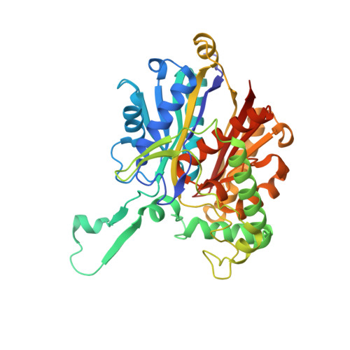
4WYS | pdb_00004wys

Crystal structure of thiolase from Escherichia coli


Experimental Data Snapshot

  • Method: X-RAY DIFFRACTION
  • Resolution: 2.10 Å
  • R-Value Free: 
    0.218 (Depositor), 0.225 (DCC) 
  • R-Value Work: 
    0.169 (Depositor), 0.184 (DCC) 
  • R-Value Observed: 
    0.172 (Depositor) 

Starting Model: experimental
View more details

wwPDB Validation 3D Report Full Report

Validation slider image for 4WYS

This is version 1.1 of the entry. See complete history. 

Literature

Redox-switch regulatory mechanism of thiolase from Clostridium acetobutylicum

Kim, S., Jang, Y.S., Ha, S.C., Ahn, J.W., Kim, E.J., Hong Lim, J., Cho, C., Shin Ryu, Y., Kuk Lee, S., Lee, S.Y., Kim, K.J.

(2015) Nat Commun 6: 8410-8410

  • DOI: https://doi.org/10.1038/ncomms9410
  • Primary Citation Related Structures: 
    4WYR, 4WYS, 4XL2, 4XL3, 4XL4

  • PubMed Abstract: 

    Thiolase is the first enzyme catalysing the condensation of two acetyl-coenzyme A (CoA) molecules to form acetoacetyl-CoA in a dedicated pathway towards the biosynthesis of n-butanol, an important solvent and biofuel. Here we elucidate the crystal structure of Clostridium acetobutylicum thiolase (CaTHL) in its reduced/oxidized states. CaTHL, unlike those from other aerobic bacteria such as Escherichia coli and Zoogloea ramegera, is regulated by the redox-switch modulation through reversible disulfide bond formation between two catalytic cysteine residues, Cys88 and Cys378. When CaTHL is overexpressed in wild-type C. acetobutylicum, butanol production is reduced due to the disturbance of acidogenic to solventogenic shift. The CaTHL(V77Q/N153Y/A286K) mutant, which is not able to form disulfide bonds, exhibits higher activity than wild-type CaTHL, and enhances butanol production upon overexpression. On the basis of these results, we suggest that CaTHL functions as a key enzyme in the regulation of the main metabolism of C. acetobutylicum through a redox-switch regulatory mechanism.


  • Organizational Affiliation: 
    • School of Life Sciences, KNU Creative BioResearch Group, Kyungpook National University, Daegu 702-701, Korea.

Macromolecule Content 

  • Total Structure Weight: 166.88 kDa 
  • Atom Count: 12,024 
  • Modeled Residue Count: 1,584 
  • Deposited Residue Count: 1,620 
  • Unique protein chains: 1

Macromolecules

Find similar proteins by:|  3D Structure
Entity ID: 1
MoleculeChains  Sequence LengthOrganismDetailsImage
Acetyl-CoA acetyltransferase
A, B, C, D
405Escherichia coli K-12Mutation(s): 0 
Gene Names: atoB, b2224, JW2218
EC: 2.3.1.9
UniProt
Find proteins for P76461 (Escherichia coli (strain K12))
Explore P76461 
Go to UniProtKB:  P76461
Entity Groups
Sequence Clusters30% Identity50% Identity70% Identity90% Identity95% Identity100% Identity
UniProt GroupP76461
Sequence Annotations
Expand
Reference Sequence

Experimental Data & Validation

Experimental Data

  • Method: X-RAY DIFFRACTION
  • Resolution: 2.10 Å
  • R-Value Free:  0.218 (Depositor), 0.225 (DCC) 
  • R-Value Work:  0.169 (Depositor), 0.184 (DCC) 
  • R-Value Observed: 0.172 (Depositor) 
Space Group: P 21 21 21
Unit Cell:
Length ( Å )Angle ( ˚ )
a = 75.007α = 90
b = 85.151β = 90
c = 269.113γ = 90
Software Package:
Software NamePurpose
MOLREPphasing
REFMACrefinement
PDB_EXTRACTdata extraction
HKL-2000data reduction
HKL-2000data scaling

Structure Validation

View Full Validation Report


Entry History 

Deposition Data

Revision History  (Full details and data files)

  • Version 1.0: 2015-10-07
    Type: Initial release
  • Version 1.1: 2023-11-08
    Changes: Data collection, Database references, Derived calculations, Refinement description