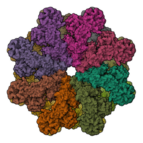
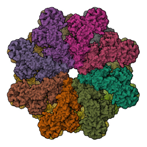
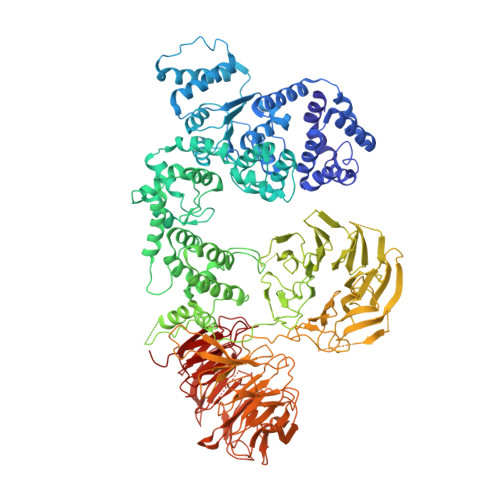
4V4L

Structure of the Drosophila apoptosome


Experimental Data Snapshot

  • Method: ELECTRON MICROSCOPY
  • Resolution: 6.90 Å
  • Aggregation State: PARTICLE 
  • Reconstruction Method: SINGLE PARTICLE 

wwPDB Validation   3D Report Full Report


This is version 1.4 of the entry. See complete history


Literature

Structure of the Drosophila apoptosome at 6.9 angstrom resolution

Yuan, S.Yu, X.Topf, M.Dorstyn, L.Kumar, S.Ludtke, S.J.Akey, C.W.

(2011) Structure 19: 128-140

  • DOI: https://doi.org/10.1016/j.str.2010.10.009
  • Primary Citation of Related Structures:  
    4V4L

  • PubMed Abstract: 

    The Drosophila Apaf-1 related killer forms an apoptosome in the intrinsic cell death pathway. In this study we show that Dark forms a single ring when initiator procaspases are bound. This Dark-Dronc complex cleaves DrICE efficiently; hence, a single ring represents the Drosophila apoptosome. We then determined the 3D structure of a double ring at ∼6.9 Å resolution and created a model of the apoptosome. Subunit interactions in the Dark complex are similar to those in Apaf-1 and CED-4 apoptosomes, but there are significant differences. In particular, Dark has "lost" a loop in the nucleotide-binding pocket, which opens a path for possible dATP exchange in the apoptosome. In addition, caspase recruitment domains (CARDs) form a crown on the central hub of the Dark apoptosome. This CARD geometry suggests that conformational changes will be required to form active Dark-Dronc complexes. When taken together, these data provide insights into apoptosome structure, function, and evolution.


  • Organizational Affiliation

    Department of Physiology and Biophysics, Boston University School of Medicine, 700 Albany Street, Boston, MA 02118, USA.


Macromolecules
Find similar proteins by:  (by identity cutoff)  |  3D Structure
Entity ID: 1
MoleculeChains Sequence LengthOrganismDetailsImage
Apaf-1 related killer DARK
A, B, C, D, E
A, B, C, D, E, F, G, H, I, J, K, L, M, N, O, P
1,221Drosophila melanogasterMutation(s): 0 
Gene Names: ArkCG6829dapaf-1LDmel_CG6829Hac1
UniProt
Find proteins for Q7KLI1 (Drosophila melanogaster)
Explore Q7KLI1 
Go to UniProtKB:  Q7KLI1
Entity Groups  
Sequence Clusters30% Identity50% Identity70% Identity90% Identity95% Identity100% Identity
UniProt GroupQ7KLI1
Sequence Annotations
Expand
  • Reference Sequence
Small Molecules
Ligands 2 Unique
IDChains Name / Formula / InChI Key2D Diagram3D Interactions
DTP
Query on DTP

Download Ideal Coordinates CCD File 
BA [auth F]
DA [auth G]
FA [auth H]
HA [auth I]
JA [auth J]
BA [auth F],
DA [auth G],
FA [auth H],
HA [auth I],
JA [auth J],
LA [auth K],
NA [auth L],
PA [auth M],
R [auth A],
RA [auth N],
T [auth B],
TA [auth O],
V [auth C],
VA [auth P],
X [auth D],
Z [auth E]
2'-DEOXYADENOSINE 5'-TRIPHOSPHATE
C10 H16 N5 O12 P3
SUYVUBYJARFZHO-RRKCRQDMSA-N
MG
Query on MG

Download Ideal Coordinates CCD File 
AA [auth F]
CA [auth G]
EA [auth H]
GA [auth I]
IA [auth J]
AA [auth F],
CA [auth G],
EA [auth H],
GA [auth I],
IA [auth J],
KA [auth K],
MA [auth L],
OA [auth M],
Q [auth A],
QA [auth N],
S [auth B],
SA [auth O],
U [auth C],
UA [auth P],
W [auth D],
Y [auth E]
MAGNESIUM ION
Mg
JLVVSXFLKOJNIY-UHFFFAOYSA-N
Experimental Data & Validation

Experimental Data

  • Method: ELECTRON MICROSCOPY
  • Resolution: 6.90 Å
  • Aggregation State: PARTICLE 
  • Reconstruction Method: SINGLE PARTICLE 
EM Software:
TaskSoftware PackageVersion
RECONSTRUCTIONEMAN2

Structure Validation

View Full Validation Report


Entry History 

Deposition Data

Revision History  (Full details and data files)

  • Version 1.0: 2014-07-09
    Type: Initial release
  • Version 1.1: 2014-12-10
    Changes: Other
  • Version 1.2: 2018-07-18
    Changes: Data collection, Database references
  • Version 1.3: 2019-12-18
    Changes: Database references, Other
  • Version 1.4: 2024-02-28
    Changes: Data collection, Database references, Derived calculations