4UI2 | pdb_00004ui2

Crystal structure of the ternary RGMB-BMP2-NEO1 complex


Experimental Data Snapshot

  • Method: X-RAY DIFFRACTION
  • Resolution: 3.15 Å
  • R-Value Free: 
    0.236 (Depositor), 0.240 (DCC) 
  • R-Value Work: 
    0.196 (Depositor), 0.203 (DCC) 
  • R-Value Observed: 
    0.198 (Depositor) 

wwPDB Validation 3D Report Full Report

Validation slider image for 4UI2

Ligand Structure Quality Assessment 


This is version 3.0 of the entry. See complete history

Literature

Repulsive Guidance Molecule is a Structural Bridge between Neogenin and Bone Morphogenetic Protein.

Healey, E.G.Bishop, B.Elegheert, J.Bell, C.H.Padilla-Parra, S.Siebold, C.

(2015) Nat Struct Mol Biol 22: 458

  • DOI: https://doi.org/10.1038/nsmb.3016
  • Primary Citation Related Structures: 
    4UHY, 4UHZ, 4UI0, 4UI1, 4UI2

  • PubMed Abstract: 

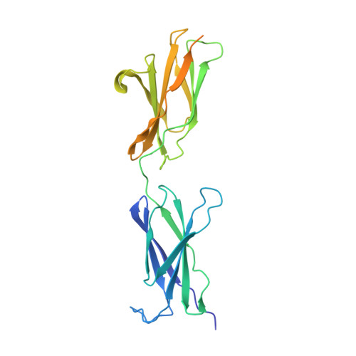
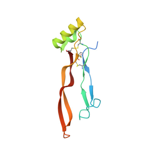
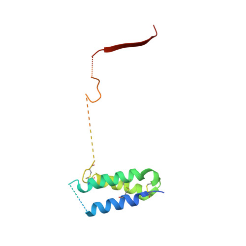
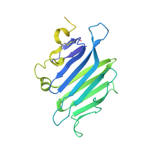
    Repulsive guidance molecules (RGMs) control crucial processes including cell motility, adhesion, immune-cell regulation and systemic iron metabolism. RGMs signal via the neogenin (NEO1) and the bone morphogenetic protein (BMP) pathways. Here, we report crystal structures of the N-terminal domains of all human RGM family members in complex with the BMP ligand BMP2, revealing a new protein fold and a conserved BMP-binding mode. Our structural and functional data suggest a pH-linked mechanism for RGM-activated BMP signaling and offer a rationale for RGM mutations causing juvenile hemochromatosis. We also determined the crystal structure of the ternary BMP2-RGM-NEO1 complex, which, along with solution scattering and live-cell super-resolution fluorescence microscopy, indicates BMP-induced clustering of the RGM-NEO1 complex. Our results show how RGM acts as the central hub that links BMP and NEO1 and physically connects these fundamental signaling pathways.


  • Organizational Affiliation
    • Division of Structural Biology, Wellcome Trust Centre for Human Genetics, University of Oxford, Oxford, UK.

Macromolecule Content 

  • Total Structure Weight: 84.51 kDa 
  • Atom Count: 4,311 
  • Modeled Residue Count: 541 
  • Deposited Residue Count: 751 
  • Unique protein chains: 4

Macromolecules

Find similar proteins by:|  3D Structure
Entity ID: 1
MoleculeChains  Sequence LengthOrganismDetailsImage
NEOGENIN264Homo sapiensMutation(s): 0 
UniProt & NIH Common Fund Data Resources
Find proteins for Q92859 (Homo sapiens)
Explore Q92859 
Go to UniProtKB:  Q92859
PHAROS:  Q92859
GTEx:  ENSG00000067141 
Entity Groups
Sequence Clusters30% Identity50% Identity70% Identity90% Identity95% Identity100% Identity
UniProt GroupQ92859
Glycosylation
Glycosylation Sites: 1Go to GlyGen: Q92859-1
Sequence Annotations
Expand
Reference Sequence
Find similar proteins by:|  3D Structure
Entity ID: 2
MoleculeChains  Sequence LengthOrganismDetailsImage
BONE MORPHOGENETIC PROTEIN 2, BMP2114Homo sapiensMutation(s): 0 
UniProt & NIH Common Fund Data Resources
Find proteins for P12643 (Homo sapiens)
Explore P12643 
Go to UniProtKB:  P12643
PHAROS:  P12643
GTEx:  ENSG00000125845 
Entity Groups
Sequence Clusters30% Identity50% Identity70% Identity90% Identity95% Identity100% Identity
UniProt GroupP12643
Sequence Annotations
Expand
Reference Sequence
Find similar proteins by:|  3D Structure
Entity ID: 3
MoleculeChains  Sequence LengthOrganismDetailsImage
REPULSIVE GUIDANCE MOLECULE C, RGMC, HEMOJUVELIN122Homo sapiensMutation(s): 0 
UniProt & NIH Common Fund Data Resources
Find proteins for Q6NW40 (Homo sapiens)
Explore Q6NW40 
Go to UniProtKB:  Q6NW40
PHAROS:  Q6NW40
GTEx:  ENSG00000174136 
Entity Groups
Sequence Clusters30% Identity50% Identity70% Identity90% Identity95% Identity100% Identity
UniProt GroupQ6NW40
Sequence Annotations
Expand
Reference Sequence
Find similar proteins by:|  3D Structure
Entity ID: 4
MoleculeChains  Sequence LengthOrganismDetailsImage
REPULSIVE GUIDANCE MOLECULE C, RGMC, HEMOJUVELIN251Homo sapiensMutation(s): 0 
UniProt & NIH Common Fund Data Resources
Find proteins for Q6NW40 (Homo sapiens)
Explore Q6NW40 
Go to UniProtKB:  Q6NW40
PHAROS:  Q6NW40
GTEx:  ENSG00000174136 
Entity Groups
Sequence Clusters30% Identity50% Identity70% Identity90% Identity95% Identity100% Identity
UniProt GroupQ6NW40
Sequence Annotations
Expand
Reference Sequence

Oligosaccharides

Help  
Entity ID: 5
MoleculeChains Length2D Diagram GlycosylationD Interactions
beta-D-mannopyranose-(1-4)-2-acetamido-2-deoxy-beta-D-glucopyranose-(1-4)-2-acetamido-2-deoxy-beta-D-glucopyranose
E
3N-Glycosylation
Glycosylation Resources
GlyTouCan: G15407YE
GlyCosmos: G15407YE
GlyGen: G15407YE

Experimental Data & Validation

Experimental Data

  • Method: X-RAY DIFFRACTION
  • Resolution: 3.15 Å
  • R-Value Free:  0.236 (Depositor), 0.240 (DCC) 
  • R-Value Work:  0.196 (Depositor), 0.203 (DCC) 
  • R-Value Observed: 0.198 (Depositor) 
Space Group: P 43 21 2
Unit Cell:
Length ( Å )Angle ( ˚ )
a = 120.08α = 90
b = 120.08β = 90
c = 204.09γ = 90
Software Package:
Software NamePurpose
REFMACrefinement
XDSdata reduction
SCALAdata scaling
PHASERphasing

Structure Validation

View Full Validation Report


Ligand Structure Quality Assessment 


Entry History 

Deposition Data

Revision History  (Full details and data files)

  • Version 1.0: 2015-05-06
    Type: Initial release
  • Version 1.1: 2015-05-20
    Changes: Database references
  • Version 1.2: 2015-06-17
    Changes: Database references
  • Version 1.3: 2018-06-20
    Changes: Advisory, Data collection, Derived calculations
  • Version 2.0: 2020-07-29
    Type: Remediation
    Reason: Carbohydrate remediation
    Changes: Advisory, Atomic model, Data collection, Derived calculations, Other, Structure summary
  • Version 2.1: 2025-04-09
    Changes: Data collection, Database references, Structure summary
  • Version 3.0: 2025-10-01
    Changes: Advisory, Atomic model, Data collection, Derived calculations, Structure summary