4U06 | pdb_00004u06

Structure of Leptospira interrogans LRR protein LIC10831


Experimental Data Snapshot

  • Method: X-RAY DIFFRACTION
  • Resolution: 1.90 Å
  • R-Value Free: 
    0.235 (Depositor), 0.245 (DCC) 
  • R-Value Work: 
    0.201 (Depositor), 0.206 (DCC) 
  • R-Value Observed: 
    0.203 (Depositor) 

wwPDB Validation 3D Report Full Report

Validation slider image for 4U06

This is version 1.2 of the entry. See complete history

Literature

Structural characterization of a novel subfamily of leucine-rich repeat proteins from the human pathogen Leptospira interrogans.

Miras, I.Saul, F.Nowakowski, M.Weber, P.Haouz, A.Shepard, W.Picardeau, M.

(2015) Acta Crystallogr D Biol Crystallogr 71: 1351-1359

  • DOI: https://doi.org/10.1107/S139900471500704X
  • Primary Citation Related Structures: 
    4TZH, 4U06, 4U08, 4U09

  • PubMed Abstract: 

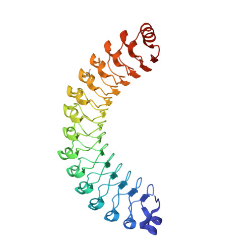
    Pathogenic Leptospira spp. are the agents of leptospirosis, an emerging zoonotic disease. Analyses of Leptospira genomes have shown that the pathogenic leptospires (but not the saprophytes) possess a large number of genes encoding proteins containing leucine-rich repeat (LRR) domains. In other pathogenic bacteria, proteins with LRR domains have been shown to be involved in mediating host-cell attachment and invasion, but their functions remain unknown in Leptospira. To gain insight into the potential function of leptospiral LRR proteins, the crystal structures of four LRR proteins that represent a novel subfamily with consecutive stretches of a 23-amino-acid LRR repeat motif have been solved. The four proteins analyzed adopt the characteristic α/β-solenoid horseshoe fold. The exposed residues of the inner concave surfaces of the solenoid, which constitute a putative functional binding site, are not conserved. The various leptospiral LRR proteins could therefore recognize distinct structural motifs of different host proteins and thus serve separate and complementary functions in the physiology of these bacteria.


  • Organizational Affiliation
    • Institut Pasteur, Plate-forme de Cristallographie, CNRS-UMR 3528, Paris, France.

Macromolecule Content 

  • Total Structure Weight: 40.78 kDa 
  • Atom Count: 3,274 
  • Modeled Residue Count: 344 
  • Deposited Residue Count: 344 
  • Unique protein chains: 1

Macromolecules

Find similar proteins by:|  3D Structure
Entity ID: 1
MoleculeChains  Sequence LengthOrganismDetailsImage
LIC10831344Leptospira interrogans serovar Copenhageni str. Fiocruz L1-130Mutation(s): 2 
Gene Names: LIC_10831
UniProt
Find proteins for Q72U33 (Leptospira interrogans serogroup Icterohaemorrhagiae serovar copenhageni (strain Fiocruz L1-130))
Explore Q72U33 
Go to UniProtKB:  Q72U33
Entity Groups
Sequence Clusters30% Identity50% Identity70% Identity90% Identity95% Identity100% Identity
UniProt GroupQ72U33
Sequence Annotations
Expand
Reference Sequence

Small Molecules

Experimental Data & Validation

Experimental Data

  • Method: X-RAY DIFFRACTION
  • Resolution: 1.90 Å
  • R-Value Free:  0.235 (Depositor), 0.245 (DCC) 
  • R-Value Work:  0.201 (Depositor), 0.206 (DCC) 
  • R-Value Observed: 0.203 (Depositor) 
Space Group: P 42 21 2
Unit Cell:
Length ( Å )Angle ( ˚ )
a = 122.3α = 90
b = 122.3β = 90
c = 58.2γ = 90
Software Package:
Software NamePurpose
BUSTERrefinement
XDSdata reduction
SCALAdata scaling
SHELXDEphasing

Structure Validation

View Full Validation Report


Entry History 

Deposition Data

Revision History  (Full details and data files)

  • Version 1.0: 2015-06-03
    Type: Initial release
  • Version 1.1: 2015-06-17
    Changes: Database references
  • Version 1.2: 2024-11-13
    Changes: Data collection, Database references, Derived calculations, Structure summary