4TRG

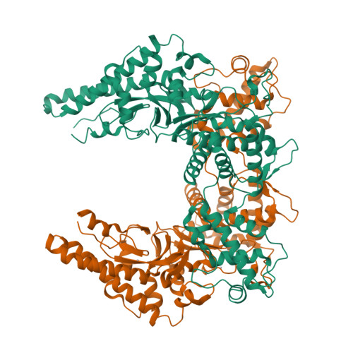
the SNL domain of SidC


Experimental Data Snapshot

  • Method: X-RAY DIFFRACTION
  • Resolution: 2.59 Å
  • R-Value Free: 0.231 
  • R-Value Work: 0.189 
  • R-Value Observed: 0.191 

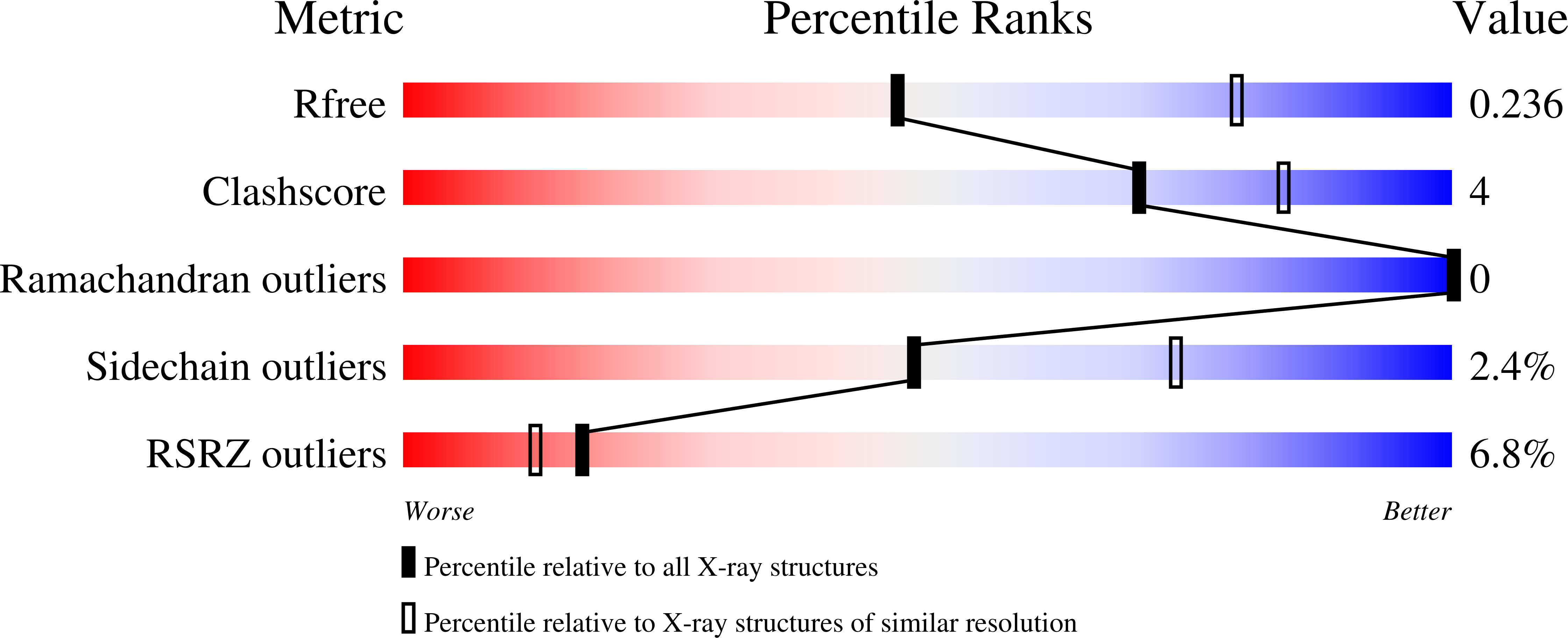
wwPDB Validation   3D Report Full Report


This is version 1.8 of the entry. See complete history


Literature

The Legionella effector SidC defines a unique family of ubiquitin ligases important for bacterial phagosomal remodeling.

Hsu, F.Luo, X.Qiu, J.Teng, Y.B.Jin, J.Smolka, M.B.Luo, Z.Q.Mao, Y.

(2014) Proc Natl Acad Sci U S A 111: 10538-10543

  • DOI: https://doi.org/10.1073/pnas.1402605111
  • Primary Citation of Related Structures:  
    4TRG, 4TRH

  • PubMed Abstract: 

    The activity of proteins delivered into host cells by the Dot/Icm injection apparatus allows Legionella pneumophila to establish a niche called the Legionella-containing vacuole (LCV), which is permissive for intracellular bacterial propagation. Among these proteins, substrate of Icm/Dot transporter (SidC) anchors to the cytoplasmic surface of the LCV and is important for the recruitment of host endoplasmic reticulum (ER) proteins to this organelle. However, the biochemical function underlying this activity is unknown. Here, we determined the structure of the N-terminal domain of SidC, which has no structural homology to any protein. Sequence homology analysis revealed a potential canonical catalytic triad formed by Cys46, His444, and Asp446 on the surface of SidC. Unexpectedly, we found that SidC is an E3 ubiquitin ligase that uses the C-H-D triad to catalyze the formation of high-molecular-weight polyubiquitin chains through multiple ubiquitin lysine residues. A C46A mutation completely abolished the E3 ligase activity and the ability of the protein to recruit host ER proteins as well as polyubiquitin conjugates to the LCV. Thus, SidC represents a unique E3 ubiquitin ligase family important for phagosomal membrane remodeling by L. pneumophila.


  • Organizational Affiliation

    Weill Institute for Cell and Molecular Biology and Department of Molecular Biology and Genetics, Cornell University, Ithaca, NY 14853;


Macromolecules
Find similar proteins by:  (by identity cutoff)  |  3D Structure
Entity ID: 1
MoleculeChains Sequence LengthOrganismDetailsImage
SidC
A, B
542Legionella pneumophilaMutation(s): 0 
UniProt
Find proteins for Q6RCR4 (Legionella pneumophila)
Explore Q6RCR4 
Go to UniProtKB:  Q6RCR4
Entity Groups  
Sequence Clusters30% Identity50% Identity70% Identity90% Identity95% Identity100% Identity
UniProt GroupQ6RCR4
Sequence Annotations
Expand
  • Reference Sequence
Experimental Data & Validation

Experimental Data

  • Method: X-RAY DIFFRACTION
  • Resolution: 2.59 Å
  • R-Value Free: 0.231 
  • R-Value Work: 0.189 
  • R-Value Observed: 0.191 
  • Space Group: P 21 21 21
Unit Cell:
Length ( Å )Angle ( ˚ )
a = 61.533α = 90
b = 133.438β = 90
c = 170.249γ = 90
Software Package:
Software NamePurpose
REFMACrefinement
HKL-2000data scaling
SHELXphasing

Structure Validation

View Full Validation Report


Entry History & Funding Information

Deposition Data


Funding OrganizationLocationGrant Number
National Institutes of Health/National Institute of General Medical Sciences (NIH/NIGMS)United StatesR01-GM094347

Revision History  (Full details and data files)

  • Version 1.0: 2014-07-02
    Type: Initial release
  • Version 1.1: 2014-07-23
    Changes: Database references
  • Version 1.2: 2014-08-06
    Changes: Structure summary
  • Version 1.3: 2014-10-01
    Changes: Database references
  • Version 1.4: 2017-09-13
    Changes: Author supporting evidence, Database references, Derived calculations, Other, Source and taxonomy
  • Version 1.5: 2017-11-22
    Changes: Refinement description
  • Version 1.6: 2019-12-25
    Changes: Author supporting evidence
  • Version 1.7: 2023-12-27
    Changes: Data collection, Database references, Derived calculations, Refinement description
  • Version 1.8: 2024-10-23
    Changes: Structure summary