4R9A

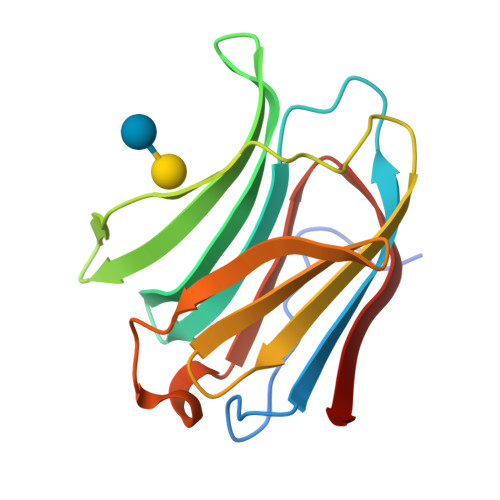
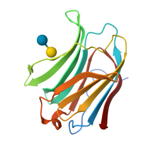
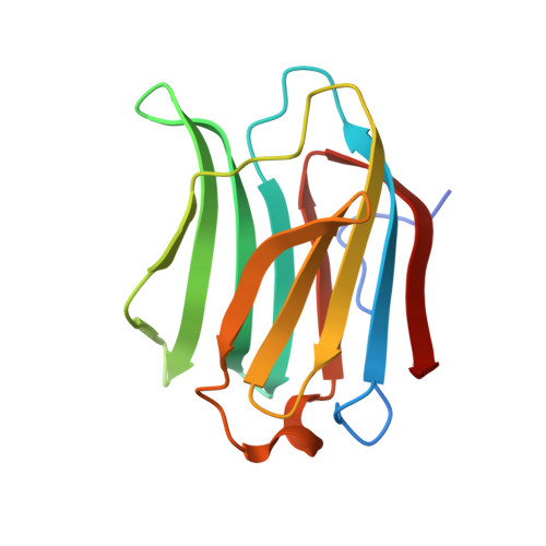
Crystal structure of Human galectin-3 CRD in complex with lactose (pH 7.0, PEG4000)


Experimental Data Snapshot

  • Method: X-RAY DIFFRACTION
  • Resolution: 1.20 Å
  • R-Value Free: 0.191 
  • R-Value Work: 0.175 
  • R-Value Observed: 0.176 

wwPDB Validation   3D Report Full Report


This is version 2.2 of the entry. See complete history


Literature

The water network in galectin-3 ligand binding site guides inhibitor design.

Su, J.Zhang, T.Wang, P.Liu, F.Tai, G.Zhou, Y.

(2015) Acta Biochim Biophys Sin (Shanghai) 47: 192-198

  • DOI: https://doi.org/10.1093/abbs/gmu132
  • Primary Citation of Related Structures:  
    4R9A, 4R9B, 4R9C, 4R9D, 4RL7

  • PubMed Abstract: 

    Galectin-3 (Gal-3) which shows affinity of β-galactosides is a cancer-related protein. Thus, it is important to understand its ligand binding mechanism and then design its specific inhibitor. It was suggested that the positions of water molecules in Gal-3 ligand-binding site could be replaced by appropriate chemical groups of ideal inhibitors. However, the reported structures of Gal-3 carbohydrate recognition domain (CRD) complexed with lactose showed that the number of water molecules are different and the water positions are inconsistent in the ligand-binding site. This study reported four high-resolution (1.24-1.19 Å) structures of Gal-3 CRD complexed with lactose, and accurately located 12 conserved water molecules in the water network of Gal-3 CRD ligand-binding site by merging these structures. These water molecules either directly stabilize the binding of Gal-3 CRD and lactose, or hold the former water molecules at the right place. In particular, water molecule 4 (W4) which only coordinates with water molecule 5 (W5) and water molecule 6 (W6) plays a key role in stabilizing galactose residue. In addition, by three-dimensional alignment of the positions of all residues, 14 flexible parts of Gal-3 CRD were found to dynamically fluctuate in the crystalline environment.


  • Organizational Affiliation

    School of Life Sciences, Northeast Normal University, Changchun 130024, China.


Macromolecules
Find similar proteins by:  (by identity cutoff)  |  3D Structure
Entity ID: 1
MoleculeChains Sequence LengthOrganismDetailsImage
Galectin-3144Homo sapiensMutation(s): 0 
Gene Names: LGALS3MAC2
UniProt & NIH Common Fund Data Resources
Find proteins for P17931 (Homo sapiens)
Explore P17931 
Go to UniProtKB:  P17931
PHAROS:  P17931
GTEx:  ENSG00000131981 
Entity Groups  
Sequence Clusters30% Identity50% Identity70% Identity90% Identity95% Identity100% Identity
UniProt GroupP17931
Sequence Annotations
Expand
  • Reference Sequence
Oligosaccharides

Help

Entity ID: 2
MoleculeChains Length2D Diagram Glycosylation3D Interactions
beta-D-galactopyranose-(1-4)-beta-D-glucopyranose
B
2N/A
Glycosylation Resources
GlyTouCan:  G84224TW
GlyCosmos:  G84224TW
GlyGen:  G84224TW
Biologically Interesting Molecules (External Reference) 1 Unique
Experimental Data & Validation

Experimental Data

  • Method: X-RAY DIFFRACTION
  • Resolution: 1.20 Å
  • R-Value Free: 0.191 
  • R-Value Work: 0.175 
  • R-Value Observed: 0.176 
  • Space Group: P 21 21 21
Unit Cell:
Length ( Å )Angle ( ˚ )
a = 36.668α = 90
b = 57.92β = 90
c = 63.032γ = 90
Software Package:
Software NamePurpose
PHENIXrefinement

Structure Validation

View Full Validation Report


Entry History 

Deposition Data

  • Released Date: 2015-03-11 
  • Deposition Author(s): Su, J.Y.

Revision History  (Full details and data files)

  • Version 1.0: 2015-03-11
    Type: Initial release
  • Version 2.0: 2020-07-29
    Type: Remediation
    Reason: Carbohydrate remediation
    Changes: Atomic model, Data collection, Database references, Derived calculations, Non-polymer description, Structure summary
  • Version 2.1: 2022-08-24
    Changes: Database references, Structure summary
  • Version 2.2: 2024-05-29
    Changes: Data collection