4QTU | pdb_00004qtu

Structure of S. cerevisiae Bud23-Trm112 complex involved in formation of m7G1575 on 18S rRNA (SAM bound form)


Experimental Data Snapshot

  • Method: X-RAY DIFFRACTION
  • Resolution: 2.12 Å
  • R-Value Free: 
    0.229 (Depositor), 0.233 (DCC) 
  • R-Value Work: 
    0.178 (Depositor), 0.186 (DCC) 
  • R-Value Observed: 
    0.181 (Depositor) 

Starting Model: experimental
View more details

wwPDB Validation 3D Report Full Report

Validation slider image for 4QTU

Ligand Structure Quality Assessment 


This is version 1.5 of the entry. See complete history

Literature

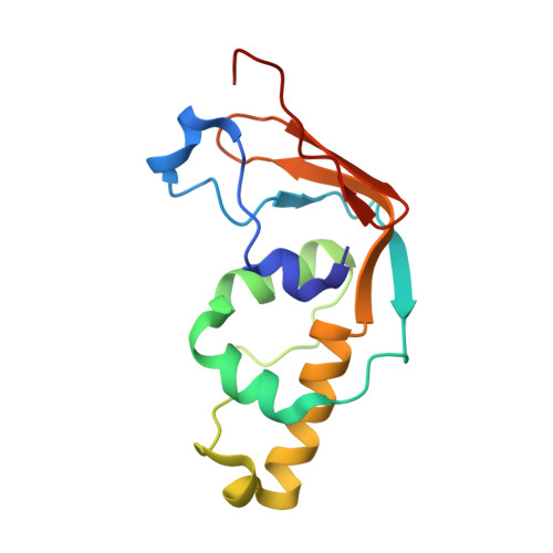
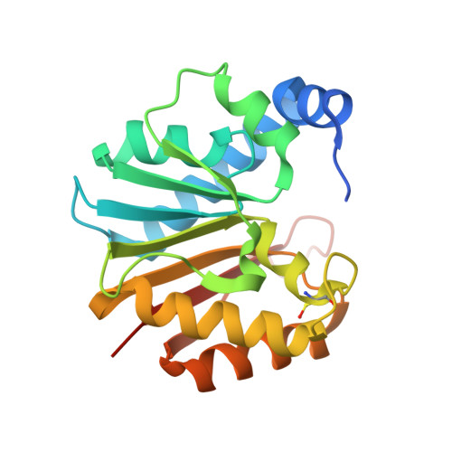
Structural and functional studies of Bud23-Trm112 reveal 18S rRNA N7-G1575 methylation occurs on late 40S precursor ribosomes.

Letoquart, J.Huvelle, E.Wacheul, L.Bourgeois, G.Zorbas, C.Graille, M.Heurgue-Hamard, V.Lafontaine, D.L.

(2014) Proc Natl Acad Sci U S A 111: E5518-E5526

  • DOI: https://doi.org/10.1073/pnas.1413089111
  • Primary Citation Related Structures: 
    4QTT, 4QTU

  • PubMed Abstract: 

    The eukaryotic small ribosomal subunit carries only four ribosomal (r) RNA methylated bases, all close to important functional sites. N(7)-methylguanosine (m(7)G) introduced at position 1575 on 18S rRNA by Bud23-Trm112 is at a ridge forming a steric block between P- and E-site tRNAs. Here we report atomic resolution structures of Bud23-Trm112 in the apo and S-adenosyl-L-methionine (SAM)-bound forms. Bud23 and Trm112 interact through formation of a β-zipper involving main-chain atoms, burying an important hydrophobic surface and stabilizing the complex. The structures revealed that the coactivator Trm112 undergoes an induced fit to accommodate its methyltransferase (MTase) partner. We report important structural similarity between the active sites of Bud23 and Coffea canephora xanthosine MTase, leading us to propose and validate experimentally a model for G1575 coordination. We identify Bud23 residues important for Bud23-Trm112 complex formation and recruitment to pre-ribosomes. We report that though Bud23-Trm112 binds precursor ribosomes at an early nucleolar stage, m(7)G methylation occurs at a late step of small subunit biogenesis, implying specifically delayed catalytic activation. Finally, we show that Bud23-Trm112 interacts directly with the box C/D snoRNA U3-associated DEAH RNA helicase Dhr1 supposedly involved in central pseudoknot formation; this suggests that Bud23-Trm112 might also contribute to controlling formation of this irreversible and dramatic structural reorganization essential to overall folding of small subunit rRNA. Our study contributes important new elements to our understanding of key molecular aspects of human ribosomopathy syndromes associated with WBSCR22 (human Bud23) malfunction.


  • Organizational Affiliation
    • Laboratoire de Biochimie, CNRS UMR 7654, Ecole Polytechnique, F-91128 Palaiseau Cedex, France;

Macromolecule Content 

  • Total Structure Weight: 78.36 kDa 
  • Atom Count: 5,256 
  • Modeled Residue Count: 639 
  • Deposited Residue Count: 686 
  • Unique protein chains: 2

Macromolecules

Find similar proteins by:|  3D Structure
Entity ID: 1
MoleculeChains  Sequence LengthOrganismDetailsImage
Multifunctional methyltransferase subunit TRM112
A, C
135Saccharomyces cerevisiae S288CMutation(s): 0 
Gene Names: N3445TRM112YNR046W
UniProt
Find proteins for P53738 (Saccharomyces cerevisiae (strain ATCC 204508 / S288c))
Explore P53738 
Go to UniProtKB:  P53738
Entity Groups
Sequence Clusters30% Identity50% Identity70% Identity90% Identity95% Identity100% Identity
UniProt GroupP53738
Sequence Annotations
Expand
Reference Sequence
Find similar proteins by:|  3D Structure
Entity ID: 2
MoleculeChains  Sequence LengthOrganismDetailsImage
Putative methyltransferase BUD23
B, D
208Saccharomyces cerevisiae S288CMutation(s): 0 
Gene Names: BUD23YCR047CYCR47C
EC: 2.1.1 (PDB Primary Data), 2.1.1.309 (UniProt)
UniProt
Find proteins for P25627 (Saccharomyces cerevisiae (strain ATCC 204508 / S288c))
Explore P25627 
Go to UniProtKB:  P25627
Entity Groups
Sequence Clusters30% Identity50% Identity70% Identity90% Identity95% Identity100% Identity
UniProt GroupP25627
Sequence Annotations
Expand
Reference Sequence

Small Molecules

Ligands 3 Unique
IDChains Name / Formula / InChI Key2D Diagram3D Interactions
SAM

Query on SAM


Download:Ideal Coordinates CCD File
L [auth B],
Z [auth D]
S-ADENOSYLMETHIONINE
C15 H22 N6 O5 S
MEFKEPWMEQBLKI-FCKMPRQPSA-N
ZN

Query on ZN


Download:Ideal Coordinates CCD File
E [auth A],
W [auth C]
ZINC ION
Zn
PTFCDOFLOPIGGS-UHFFFAOYSA-N
EDO

Query on EDO


Download:Ideal Coordinates CCD File
AA [auth D]
F [auth A]
G [auth A]
H [auth A]
I [auth A]
AA [auth D],
F [auth A],
G [auth A],
H [auth A],
I [auth A],
J [auth A],
K [auth A],
M [auth B],
N [auth B],
O [auth B],
P [auth B],
Q [auth B],
R [auth B],
S [auth B],
T [auth B],
U [auth B],
V [auth B],
X [auth C],
Y [auth C]
1,2-ETHANEDIOL
C2 H6 O2
LYCAIKOWRPUZTN-UHFFFAOYSA-N
Modified Residues  1 Unique
IDChains TypeFormula2D DiagramParent
CSO
Query on CSO
B, D
L-PEPTIDE LINKINGC3 H7 N O3 SCYS

Experimental Data & Validation

Experimental Data

  • Method: X-RAY DIFFRACTION
  • Resolution: 2.12 Å
  • R-Value Free:  0.229 (Depositor), 0.233 (DCC) 
  • R-Value Work:  0.178 (Depositor), 0.186 (DCC) 
  • R-Value Observed: 0.181 (Depositor) 
Space Group: P 1 21 1
Unit Cell:
Length ( Å )Angle ( ˚ )
a = 75.472α = 90
b = 53.733β = 94.55
c = 85.205γ = 90
Software Package:
Software NamePurpose
XDSdata scaling
SHARPphasing
PHENIXrefinement
XDSdata reduction
XSCALEdata scaling

Structure Validation

View Full Validation Report


Ligand Structure Quality Assessment 


Entry History 

Deposition Data

Revision History  (Full details and data files)

  • Version 1.0: 2014-12-24
    Type: Initial release
  • Version 1.1: 2018-01-03
    Changes: Structure summary
  • Version 1.2: 2022-08-24
    Changes: Database references, Derived calculations
  • Version 1.3: 2023-11-08
    Changes: Data collection, Refinement description
  • Version 1.4: 2023-12-06
    Changes: Data collection
  • Version 1.5: 2024-10-30
    Changes: Structure summary