Discovery of a new ATP-binding motif involved in peptidic azoline biosynthesis.
Dunbar, K.L., Chekan, J.R., Cox, C.L., Burkhart, B.J., Nair, S.K., Mitchell, D.A.(2014) Nat Chem Biol 10: 823-829
- PubMed: 25129028 Search on PubMedSearch on PubMed Central
- DOI: https://doi.org/10.1038/nchembio.1608
- Primary Citation Related Structures:
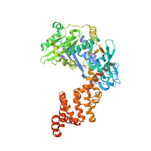
4Q84, 4Q85, 4Q86 - PubMed Abstract:
Despite intensive research, the cyclodehydratase responsible for azoline biogenesis in thiazole/oxazole-modified microcin (TOMM) natural products remains enigmatic. The collaboration of two proteins, C and D, is required for cyclodehydration. The C protein is homologous to E1 ubiquitin-activating enzymes, whereas the D protein is within the YcaO superfamily. Recent studies have demonstrated that TOMM YcaOs phosphorylate amide carbonyl oxygens to facilitate azoline formation. Here we report the X-ray crystal structure of an uncharacterized YcaO from Escherichia coli (Ec-YcaO). Ec-YcaO harbors an unprecedented fold and ATP-binding motif. This motif is conserved among TOMM YcaOs and is required for cyclodehydration. Furthermore, we demonstrate that the C protein regulates substrate binding and catalysis and that the proline-rich C terminus of the D protein is involved in C protein recognition and catalysis. This study identifies the YcaO active site and paves the way for the characterization of the numerous YcaO domains not associated with TOMM biosynthesis.
- 1] Department of Chemistry, University of Illinois at Urbana-Champaign, Urbana, Illinois, USA. [2] Institute for Genomic Biology, University of Illinois at Urbana-Champaign, Urbana, Illinois, USA. [3].
Organizational Affiliation: