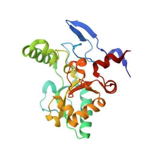
4OX3 | pdb_00004ox3

Structure of the LdcB LD-carboxypeptidase reveals the molecular basis of peptidoglycan recognition


Experimental Data Snapshot

  • Method: X-RAY DIFFRACTION
  • Resolution: 2.00 Å
  • R-Value Free: 
    0.251 (Depositor), 0.233 (DCC) 
  • R-Value Work: 
    0.205 (Depositor) 
  • R-Value Observed: 
    0.207 (Depositor) 

wwPDB Validation 3D Report Full Report

Validation slider image for 4OX3

This is version 1.6 of the entry. See complete history. 

Literature

Structure of the LdcB LD-carboxypeptidase reveals the molecular basis of peptidoglycan recognition.

Hoyland, C.N., Aldridge, C., Cleverley, R.M., Duchene, M.C., Minasov, G., Onopriyenko, O., Sidiq, K., Stogios, P.J., Anderson, W.F., Daniel, R.A., Savchenko, A., Vollmer, W., Lewis, R.J.

(2014) Structure 22: 949-960

  • DOI: https://doi.org/10.1016/j.str.2014.04.015
  • Primary Citation Related Structures: 
    4JID, 4MPH, 4OX3, 4OX5, 4OXD

  • PubMed Abstract: 

    Peptidoglycan surrounds the bacterial cytoplasmic membrane to protect the cell against osmolysis. The biosynthesis of peptidoglycan, made of glycan strands crosslinked by short peptides, is the target of antibiotics like β-lactams and glycopeptides. Nascent peptidoglycan contains pentapeptides that are trimmed by carboxypeptidases to tetra- and tripeptides. The well-characterized DD-carboxypeptidases hydrolyze the terminal D-alanine from the stem pentapeptide to produce a tetrapeptide. However, few LD-carboxypeptidases that produce tripeptides have been identified, and nothing is known about substrate specificity in these enzymes. We report biochemical properties and crystal structures of the LD-carboxypeptidases LdcB from Streptococcus pneumoniae, Bacillus anthracis, and Bacillus subtilis. The enzymes are active against bacterial cell wall tetrapeptides and adopt a zinc-carboxypeptidase fold characteristic of the LAS superfamily. We have also solved the structure of S. pneumoniae LdcB with a product mimic, elucidating the residues essential for peptidoglycan recognition and the conformational changes that occur on ligand binding.


  • Organizational Affiliation: 
    • Institute for Cell and Molecular Biosciences, Newcastle University, Newcastle upon Tyne NE2 4HH, UK.

Macromolecule Content 

  • Total Structure Weight: 25.42 kDa 
  • Atom Count: 1,912 
  • Modeled Residue Count: 214 
  • Deposited Residue Count: 219 
  • Unique protein chains: 1

Macromolecules

Find similar proteins by:|  3D Structure
Entity ID: 1
MoleculeChains  Sequence LengthOrganismDetailsImage
Putative carboxypeptidase YodJ219Bacillus subtilis subsp. subtilis str. 168Mutation(s): 0 
Gene Names: BSU19620, YodJ, yokZ
EC: 3.4
UniProt
Find proteins for O34866 (Bacillus subtilis (strain 168))
Explore O34866 
Go to UniProtKB:  O34866
Entity Groups
Sequence Clusters30% Identity50% Identity70% Identity90% Identity95% Identity100% Identity
UniProt GroupO34866
Sequence Annotations
Expand
Reference Sequence

Experimental Data & Validation

Experimental Data

  • Method: X-RAY DIFFRACTION
  • Resolution: 2.00 Å
  • R-Value Free:  0.251 (Depositor), 0.233 (DCC) 
  • R-Value Work:  0.205 (Depositor) 
  • R-Value Observed: 0.207 (Depositor) 
Space Group: P 21 21 21
Unit Cell:
Length ( Å )Angle ( ˚ )
a = 42.644α = 90
b = 53.695β = 90
c = 102.33γ = 90
Software Package:
Software NamePurpose
REFMACrefinement

Structure Validation

View Full Validation Report


Entry History 

& Funding Information

Deposition Data


Funding OrganizationLocationGrant Number
Biotechnology and Biological Sciences Research Council (BBSRC)United Kingdom--

Revision History  (Full details and data files)

  • Version 1.0: 2014-06-11
    Type: Initial release
  • Version 1.1: 2014-07-16
    Changes: Database references
  • Version 1.2: 2014-10-01
    Changes: Database references
  • Version 1.3: 2015-02-04
    Changes: Derived calculations
  • Version 1.4: 2017-09-27
    Changes: Author supporting evidence, Data collection, Database references, Derived calculations, Other, Source and taxonomy
  • Version 1.5: 2020-01-01
    Changes: Author supporting evidence
  • Version 1.6: 2023-12-27
    Changes: Data collection, Database references, Refinement description