


The structure of aquaporin
Vahedi-Faridi, A., Lodowski, D., Schenk, A., Kaptan, S., De groot, B., Walz, T., Engel, A.To be published.
Experimental Data Snapshot
wwPDB Validation 3D Report Full Report

Entity ID: 1 | |||||
|---|---|---|---|---|---|
| Molecule | Chains | Sequence Length | Organism | Details | Image |
| Aquaporin-2 | A [auth X] | 274 | Homo sapiens | Mutation(s): 1 Gene Names: AQP2 Membrane Entity: Yes | 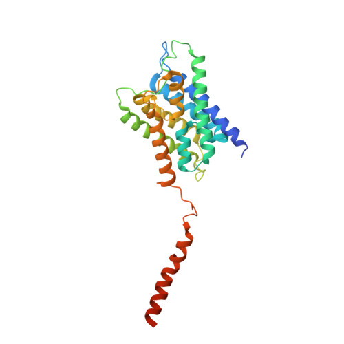 |
UniProt & NIH Common Fund Data Resources | |||||
PHAROS: P41181 GTEx: ENSG00000167580 | |||||
Entity Groups | |||||
| Sequence Clusters | 30% Identity50% Identity70% Identity90% Identity95% Identity100% Identity | ||||
| UniProt Group | P41181 | ||||
Sequence AnnotationsExpand | |||||
Reference Sequence | |||||
| Length ( Å ) | Angle ( ˚ ) |
|---|---|
| a = 95.717 | α = 90 |
| b = 95.717 | β = 90 |
| c = 79.058 | γ = 90 |
| Software Name | Purpose |
|---|---|
| ADSC | data collection |
| SOLVE | phasing |
| REFMAC | refinement |
| HKL-2000 | data reduction |
| HKL-2000 | data scaling |


