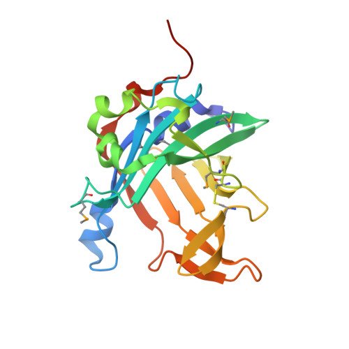
4O4S

Crystal structure of phycobiliprotein lyase CpcT complexed with phycocyanobilin (PCB)


Experimental Data Snapshot

  • Method: X-RAY DIFFRACTION
  • Resolution: 2.50 Å
  • R-Value Free: 0.248 
  • R-Value Work: 0.180 
  • R-Value Observed: 0.184 

Starting Model: experimental
View more details

wwPDB Validation   3D Report Full Report


Ligand Structure Quality Assessment 


This is version 1.6 of the entry. See complete history


Literature

Structure and Mechanism of the Phycobiliprotein Lyase CpcT.

Zhou, W.Ding, W.L.Zeng, X.L.Dong, L.L.Zhao, B.Zhou, M.Scheer, H.Zhao, K.H.Yang, X.

(2014) J Biol Chem 289: 26677-26689

  • DOI: https://doi.org/10.1074/jbc.M114.586743
  • Primary Citation of Related Structures:  
    4O4O, 4O4S

  • PubMed Abstract: 

    Pigmentation of light-harvesting phycobiliproteins of cyanobacteria requires covalent attachment of open-chain tetrapyrroles, bilins, to the apoproteins. Thioether formation via addition of a cysteine residue to the 3-ethylidene substituent of bilins is mediated by lyases. T-type lyases are responsible for attachment to Cys-155 of phycobiliprotein β-subunits. We present crystal structures of CpcT (All5339) from Nostoc (Anabaena) sp. PCC 7120 and its complex with phycocyanobilin at 1.95 and 2.50 Å resolution, respectively. CpcT forms a dimer and adopts a calyx-shaped β-barrel fold. Although the overall structure of CpcT is largely retained upon chromophore binding, arginine residues at the opening of the binding pocket undergo major rotameric rearrangements anchoring the propionate groups of phycocyanobilin. Based on the structure and mutational analysis, a reaction mechanism is proposed that accounts for chromophore stabilization and regio- and stereospecificity of the addition reaction. At the dimer interface, a loop extending from one subunit partially shields the opening of the phycocyanobilin binding pocket in the other subunit. Deletion of the loop or disruptions of the dimer interface significantly reduce CpcT lyase activity, suggesting functional relevance of the dimer. Dimerization is further enhanced by chromophore binding. The chromophore is largely buried in the dimer, but in the monomer, the 3-ethylidene group is accessible for the apophycobiliprotein, preferentially from the chromophore α-side. Asp-163 and Tyr-65 at the β- and α-face near the E-configured ethylidene group, respectively, support the acid-catalyzed nucleophilic Michael addition of cysteine 155 of the apoprotein to an N-acylimmonium intermediate proposed by Grubmayr and Wagner (Grubmayr, K., and Wagner, U. G. (1988) Monatsh. Chem. 119, 965-983).


  • Organizational Affiliation

    State Key Laboratory of Agricultural Microbiology, Huazhong Agricultural University, Wuhan 430070, China.


Macromolecules
Find similar proteins by:  (by identity cutoff)  |  3D Structure
Entity ID: 1
MoleculeChains Sequence LengthOrganismDetailsImage
Phycocyanobilin lyase CpcT207Nostoc sp. PCC 7120 = FACHB-418Mutation(s): 0 
Gene Names: cpcT1cpeT1all5339
EC: 4
UniProt
Find proteins for Q8YLF9 (Nostoc sp. (strain PCC 7120 / SAG 25.82 / UTEX 2576))
Explore Q8YLF9 
Go to UniProtKB:  Q8YLF9
Entity Groups  
Sequence Clusters30% Identity50% Identity70% Identity90% Identity95% Identity100% Identity
UniProt GroupQ8YLF9
Sequence Annotations
Expand
  • Reference Sequence
Small Molecules
Ligands 1 Unique
IDChains Name / Formula / InChI Key2D Diagram3D Interactions
CYC
Query on CYC

Download Ideal Coordinates CCD File 
M [auth A]
N [auth B]
O [auth I]
P [auth J]
Q [auth C]
M [auth A],
N [auth B],
O [auth I],
P [auth J],
Q [auth C],
R [auth D],
S [auth E],
T [auth F],
U [auth G],
V [auth H],
W [auth K],
X [auth L]
PHYCOCYANOBILIN
C33 H40 N4 O6
VXTXPYZGDQPMHK-GMXXPEQVSA-N
Modified Residues  1 Unique
IDChains TypeFormula2D DiagramParent
MSE
Query on MSE
A
B
C [auth I]
D [auth J]
E [auth C]
A,
B,
C [auth I],
D [auth J],
E [auth C],
F [auth D],
G [auth E],
H [auth F],
I [auth G],
J [auth H],
K,
L
L-PEPTIDE LINKINGC5 H11 N O2 SeMET
Experimental Data & Validation

Experimental Data

  • Method: X-RAY DIFFRACTION
  • Resolution: 2.50 Å
  • R-Value Free: 0.248 
  • R-Value Work: 0.180 
  • R-Value Observed: 0.184 
  • Space Group: P 1
Unit Cell:
Length ( Å )Angle ( ˚ )
a = 69.746α = 90.21
b = 69.601β = 90.28
c = 162.595γ = 60.12
Software Package:
Software NamePurpose
MAR345dtbdata collection
PHASERphasing
PHENIXrefinement
HKL-2000data reduction
HKL-2000data scaling

Structure Validation

View Full Validation Report


Ligand Structure Quality Assessment 


Entry History 

Deposition Data

Revision History  (Full details and data files)

  • Version 1.0: 2014-08-13
    Type: Initial release
  • Version 1.1: 2014-09-17
    Changes: Database references
  • Version 1.2: 2014-10-15
    Changes: Database references
  • Version 1.3: 2023-09-20
    Changes: Data collection, Database references, Derived calculations, Refinement description
  • Version 1.4: 2023-12-06
    Changes: Data collection
  • Version 1.5: 2024-01-10
    Changes: Derived calculations
  • Version 1.6: 2024-10-30
    Changes: Structure summary