4MYH | pdb_00004myh

Structure of the Glutathione bound mitochondrial ABC transporter, Atm1


Experimental Data Snapshot

  • Method: X-RAY DIFFRACTION
  • Resolution: 3.38 Å
  • R-Value Free: 
    0.290 (Depositor), 0.291 (DCC) 
  • R-Value Work: 
    0.247 (Depositor), 0.250 (DCC) 
  • R-Value Observed: 
    0.250 (Depositor) 

wwPDB Validation 3D Report Full Report

Validation slider image for 4MYH

Ligand Structure Quality Assessment 


This is version 1.2 of the entry. See complete history

Literature

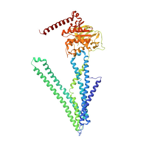
Crystal structures of nucleotide-free and glutathione-bound mitochondrial ABC transporter Atm1.

Srinivasan, V.Pierik, A.J.Lill, R.

(2014) Science 343: 1137-1140

  • DOI: https://doi.org/10.1126/science.1246729
  • Primary Citation Related Structures: 
    4MYC, 4MYH

  • PubMed Abstract: 

    The yeast mitochondrial ABC transporter Atm1, in concert with glutathione, functions in the export of a substrate required for cytosolic-nuclear iron-sulfur protein biogenesis and cellular iron regulation. Defects in the human ortholog ABCB7 cause the sideroblastic anemia XLSA/A. Here, we report the crystal structures of free and glutathione-bound Atm1 in inward-facing, open conformations at 3.06- and 3.38-angstrom resolution, respectively. The glutathione binding site includes a residue mutated in XLSA/A and is located close to the inner membrane surface in a large cavity. The two nucleotide-free adenosine 5'-triphosphate binding domains do not interact yet are kept in close vicinity through tight interaction of the two C-terminal α-helices of the Atm1 dimer. The resulting protein stabilization may be a common structural feature of all ABC exporters.


  • Organizational Affiliation
    • Institut für Zytobiologie, Philipps-Universität Marburg, Robert-Koch-Strasse 6, 35032 Marburg, Germany.

Macromolecule Content 

  • Total Structure Weight: 201.94 kDa 
  • Atom Count: 14,048 
  • Modeled Residue Count: 1,794 
  • Deposited Residue Count: 1,794 
  • Unique protein chains: 1

Macromolecules

Find similar proteins by:|  3D Structure
Entity ID: 1
MoleculeChains  Sequence LengthOrganismDetailsImage
Iron-sulfur clusters transporter ATM1, mitochondrialA,
B [auth C],
C [auth B]
598Saccharomyces cerevisiae S288CMutation(s): 0 
Gene Names: ATM1MDYYMR301CYM9952.03C
EC: 3.6.3.44 (PDB Primary Data), 7 (UniProt)
Membrane Entity: Yes 
UniProt
Find proteins for P40416 (Saccharomyces cerevisiae (strain ATCC 204508 / S288c))
Explore P40416 
Go to UniProtKB:  P40416
Entity Groups
Sequence Clusters30% Identity50% Identity70% Identity90% Identity95% Identity100% Identity
UniProt GroupP40416
Sequence Annotations
Expand
Reference Sequence

Small Molecules

Ligands 1 Unique
IDChains Name / Formula / InChI Key2D Diagram3D Interactions
GSH

Query on GSH


Download:Ideal Coordinates CCD File
D [auth B]Glutathione
C10 H17 N3 O6 S
RWSXRVCMGQZWBV-WDSKDSINSA-N

Biologically Interesting Molecules (External Reference) 

1 Unique

Experimental Data & Validation

Experimental Data

  • Method: X-RAY DIFFRACTION
  • Resolution: 3.38 Å
  • R-Value Free:  0.290 (Depositor), 0.291 (DCC) 
  • R-Value Work:  0.247 (Depositor), 0.250 (DCC) 
  • R-Value Observed: 0.250 (Depositor) 
Space Group: P 65 2 2
Unit Cell:
Length ( Å )Angle ( ˚ )
a = 157.686α = 90
b = 157.686β = 90
c = 523.29γ = 120
Software Package:
Software NamePurpose
SHELXCDphasing
PHENIXmodel building
PHENIXrefinement
XDSdata reduction
SCALAdata scaling
PHENIXphasing

Structure Validation

View Full Validation Report


Ligand Structure Quality Assessment 


Entry History 

Deposition Data

Revision History  (Full details and data files)

  • Version 1.0: 2014-03-26
    Type: Initial release
  • Version 1.1: 2017-11-15
    Changes: Refinement description
  • Version 1.2: 2024-02-28
    Changes: Data collection, Database references, Derived calculations