4MVJ | pdb_00004mvj

2.85 Angstrom Resolution Crystal Structure of Glyceraldehyde 3-phosphate Dehydrogenase A (gapA) from Escherichia coli Modified by Acetyl Phosphate.


Experimental Data Snapshot

  • Method: X-RAY DIFFRACTION
  • Resolution: 2.85 Å
  • R-Value Free: 
    0.224 (Depositor), 0.221 (DCC) 
  • R-Value Work: 
    0.186 (Depositor), 0.184 (DCC) 
  • R-Value Observed: 
    0.188 (Depositor) 

Starting Model: experimental
View more details

wwPDB Validation 3D Report Full Report

Validation slider image for 4MVJ

Ligand Structure Quality Assessment 


This is version 1.5 of the entry. See complete history

Literature

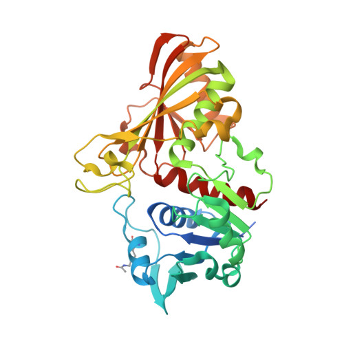
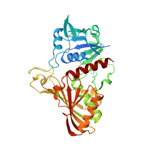
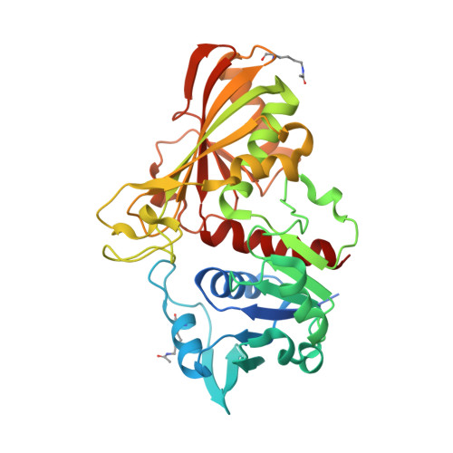
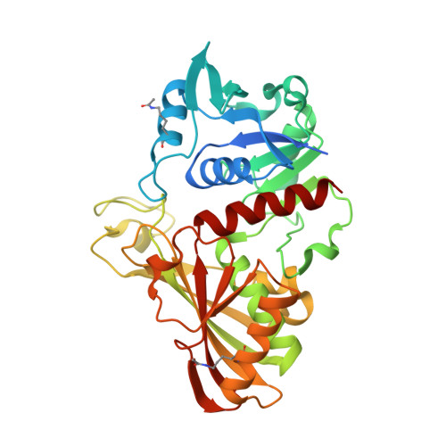
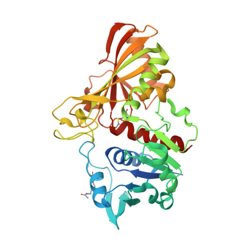
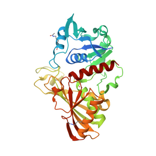
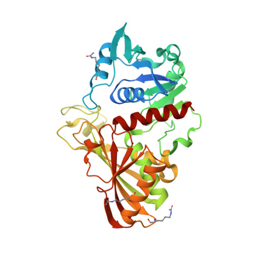
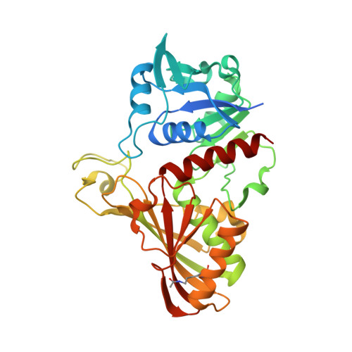
Structural, kinetic and proteomic characterization of acetyl phosphate-dependent bacterial protein acetylation.

Kuhn, M.L.Zemaitaitis, B.Hu, L.I.Sahu, A.Sorensen, D.Minasov, G.Lima, B.P.Scholle, M.Mrksich, M.Anderson, W.F.Gibson, B.W.Schilling, B.Wolfe, A.J.

(2014) PLoS One 9: e94816-e94816

  • DOI: https://doi.org/10.1371/journal.pone.0094816
  • Primary Citation Related Structures: 
    4K6A, 4MVA, 4MVJ

  • PubMed Abstract: 

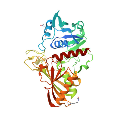
    The emerging view of Nε-lysine acetylation in eukaryotes is of a relatively abundant post-translational modification (PTM) that has a major impact on the function, structure, stability and/or location of thousands of proteins involved in diverse cellular processes. This PTM is typically considered to arise by the donation of the acetyl group from acetyl-coenzyme A (acCoA) to the ε-amino group of a lysine residue that is reversibly catalyzed by lysine acetyltransferases and deacetylases. Here, we provide genetic, mass spectrometric, biochemical and structural evidence that Nε-lysine acetylation is an equally abundant and important PTM in bacteria. Applying a recently developed, label-free and global mass spectrometric approach to an isogenic set of mutants, we detected acetylation of thousands of lysine residues on hundreds of Escherichia coli proteins that participate in diverse and often essential cellular processes, including translation, transcription and central metabolism. Many of these acetylations were regulated in an acetyl phosphate (acP)-dependent manner, providing compelling evidence for a recently reported mechanism of bacterial Nε-lysine acetylation. These mass spectrometric data, coupled with observations made by crystallography, biochemistry, and additional mass spectrometry showed that this acP-dependent acetylation is both non-enzymatic and specific, with specificity determined by the accessibility, reactivity and three-dimensional microenvironment of the target lysine. Crystallographic evidence shows acP can bind to proteins in active sites and cofactor binding sites, but also potentially anywhere molecules with a phosphate moiety could bind. Finally, we provide evidence that acP-dependent acetylation can impact the function of critical enzymes, including glyceraldehyde-3-phosphate dehydrogenase, triosephosphate isomerase, and RNA polymerase.


  • Organizational Affiliation
    • Center for Structural Genomics of Infectious Diseases, Department of Molecular Pharmacology and Biological Chemistry, Northwestern University Feinberg School of Medicine, Chicago, Illinois, United States of America.

Macromolecule Content 

  • Total Structure Weight: 623.96 kDa 
  • Atom Count: 41,155 
  • Modeled Residue Count: 5,294 
  • Deposited Residue Count: 5,680 
  • Unique protein chains: 9

Macromolecules

Find similar proteins by:|  3D Structure
Entity ID: 1
MoleculeChains  Sequence LengthOrganismDetailsImage
Glyceraldehyde-3-phosphate dehydrogenase A
A, B, C, K, N
355Escherichia coli DH1Mutation(s): 0 
Gene Names: ECDH1ME8569_1723EcDH1_1863gapA
EC: 1.2.1.12
Find similar proteins by:|  3D Structure
Entity ID: 2
MoleculeChains  Sequence LengthOrganismDetailsImage
Glyceraldehyde-3-phosphate dehydrogenase A
D, L
355Escherichia coli DH1Mutation(s): 0 
Gene Names: ECDH1ME8569_1723EcDH1_1863gapA
EC: 1.2.1.12
Find similar proteins by:|  3D Structure
Entity ID: 3
MoleculeChains  Sequence LengthOrganismDetailsImage
Glyceraldehyde-3-phosphate Dehydrogenase A355Escherichia coli DH1Mutation(s): 0 
Gene Names: ECDH1ME8569_1723EcDH1_1863gapA
EC: 1.2.1.12
Find similar proteins by:|  3D Structure
Entity ID: 4
MoleculeChains  Sequence LengthOrganismDetailsImage
Glyceraldehyde-3-phosphate Dehydrogenase A
F, J, O
355Escherichia coli DH1Mutation(s): 0 
Gene Names: ECDH1ME8569_1723EcDH1_1863gapA
EC: 1.2.1.12
Find similar proteins by:|  3D Structure
Entity ID: 5
MoleculeChains  Sequence LengthOrganismDetailsImage
Glyceraldehyde-3-phosphate Dehydrogenase A355Escherichia coli DH1Mutation(s): 0 
Gene Names: ECDH1ME8569_1723EcDH1_1863gapA
EC: 1.2.1.12
Find similar proteins by:|  3D Structure
Entity ID: 6
MoleculeChains  Sequence LengthOrganismDetailsImage
Glyceraldehyde-3-phosphate Dehydrogenase A355Escherichia coli DH1Mutation(s): 0 
Gene Names: ECDH1ME8569_1723EcDH1_1863gapA
EC: 1.2.1.12
Find similar proteins by:|  3D Structure
Entity ID: 7
MoleculeChains  Sequence LengthOrganismDetailsImage
Glyceraldehyde-3-phosphate Dehydrogenase A355Escherichia coli DH1Mutation(s): 0 
Gene Names: ECDH1ME8569_1723EcDH1_1863gapA
EC: 1.2.1.12
Find similar proteins by:|  3D Structure
Entity ID: 8
MoleculeChains  Sequence LengthOrganismDetailsImage
Glyceraldehyde-3-phosphate Dehydrogenase A355Escherichia coli DH1Mutation(s): 0 
Gene Names: ECDH1ME8569_1723EcDH1_1863gapA
EC: 1.2.1.12
Find similar proteins by:|  3D Structure
Entity ID: 9
MoleculeChains  Sequence LengthOrganismDetailsImage
Glyceraldehyde-3-phosphate Dehydrogenase A355Escherichia coli DH1Mutation(s): 0 
Gene Names: ECDH1ME8569_1723EcDH1_1863gapA
EC: 1.2.1.12

Small Molecules

Ligands 10 Unique
IDChains Name / Formula / InChI Key2D Diagram3D Interactions
NAD

Query on NAD


Download:Ideal Coordinates CCD File
BD [auth P]
HC [auth K]
JA [auth C]
QA [auth D]
WB [auth I]
BD [auth P],
HC [auth K],
JA [auth C],
QA [auth D],
WB [auth I],
YC [auth O],
ZA [auth E]
NICOTINAMIDE-ADENINE-DINUCLEOTIDE
C21 H27 N7 O14 P2
BAWFJGJZGIEFAR-NNYOXOHSSA-N
PG4

Query on PG4


Download:Ideal Coordinates CCD File
FC [auth J]TETRAETHYLENE GLYCOL
C8 H18 O5
UWHCKJMYHZGTIT-UHFFFAOYSA-N
POP

Query on POP


Download:Ideal Coordinates CCD File
GC [auth J]
IA [auth B]
JB [auth F]
PB [auth G]
VC [auth M]
GC [auth J],
IA [auth B],
JB [auth F],
PB [auth G],
VC [auth M],
Y [auth A]
PYROPHOSPHATE 2-
H2 O7 P2
XPPKVPWEQAFLFU-UHFFFAOYSA-L
PGE

Query on PGE


Download:Ideal Coordinates CCD File
UB [auth H]TRIETHYLENE GLYCOL
C6 H14 O4
ZIBGPFATKBEMQZ-UHFFFAOYSA-N
UVW

Query on UVW


Download:Ideal Coordinates CCD File
UC [auth M]ACETYLPHOSPHATE
C2 H5 O5 P
LIPOUNRJVLNBCD-UHFFFAOYSA-N
PEG

Query on PEG


Download:Ideal Coordinates CCD File
VB [auth H]DI(HYDROXYETHYL)ETHER
C4 H10 O3
MTHSVFCYNBDYFN-UHFFFAOYSA-N
PO4

Query on PO4


Download:Ideal Coordinates CCD File
AC [auth I]
DC [auth J]
EC [auth J]
ED [auth P]
FD [auth P]
AC [auth I],
DC [auth J],
EC [auth J],
ED [auth P],
FD [auth P],
GB [auth E],
HA [auth B],
IB [auth F],
QC [auth L],
SB [auth H],
V [auth A],
W [auth A],
X [auth A],
XA [auth D],
YA [auth D]
PHOSPHATE ION
O4 P
NBIIXXVUZAFLBC-UHFFFAOYSA-K
ACT

Query on ACT


Download:Ideal Coordinates CCD File
TB [auth H]ACETATE ION
C2 H3 O2
QTBSBXVTEAMEQO-UHFFFAOYSA-M
CL

Query on CL


Download:Ideal Coordinates CCD File
AD [auth O]
BA [auth B]
BB [auth E]
BC [auth J]
CA [auth B]
AD [auth O],
BA [auth B],
BB [auth E],
BC [auth J],
CA [auth B],
CB [auth E],
CC [auth J],
CD [auth P],
DA [auth B],
DB [auth E],
DD [auth P],
EA [auth B],
EB [auth E],
FA [auth B],
FB [auth E],
GA [auth B],
HB [auth F],
JC [auth K],
KC [auth K],
LA [auth C],
LB [auth G],
LC [auth K],
MA [auth C],
MB [auth G],
MC [auth K],
NA [auth C],
NB [auth G],
NC [auth K],
OA [auth C],
OB [auth G],
OC [auth K],
PA [auth C],
PC [auth L],
R [auth A],
RB [auth H],
S [auth A],
SA [auth D],
SC [auth M],
T [auth A],
TA [auth D],
TC [auth M],
U [auth A],
UA [auth D],
VA [auth D],
WA [auth D],
XC [auth N],
YB [auth I],
ZB [auth I],
ZC [auth O]
CHLORIDE ION
Cl
VEXZGXHMUGYJMC-UHFFFAOYSA-M
NA

Query on NA


Download:Ideal Coordinates CCD File
AA [auth B]
AB [auth E]
IC [auth K]
KA [auth C]
KB [auth G]
AA [auth B],
AB [auth E],
IC [auth K],
KA [auth C],
KB [auth G],
Q [auth A],
QB [auth H],
RA [auth D],
RC [auth M],
WC [auth N],
XB [auth I],
Z [auth B]
SODIUM ION
Na
FKNQFGJONOIPTF-UHFFFAOYSA-N
Modified Residues  2 Unique
IDChains TypeFormula2D DiagramParent
ALY
Query on ALY
A, B, C, K, N
L-PEPTIDE LINKINGC8 H16 N2 O3LYS
SCY
Query on SCY
G
L-PEPTIDE LINKINGC5 H9 N O3 SCYS

Experimental Data & Validation

Experimental Data

  • Method: X-RAY DIFFRACTION
  • Resolution: 2.85 Å
  • R-Value Free:  0.224 (Depositor), 0.221 (DCC) 
  • R-Value Work:  0.186 (Depositor), 0.184 (DCC) 
  • R-Value Observed: 0.188 (Depositor) 
Space Group: P 1 21 1
Unit Cell:
Length ( Å )Angle ( ˚ )
a = 145.879α = 90
b = 69.688β = 98.8
c = 271.923γ = 90
Software Package:
Software NamePurpose
Blu-Icedata collection
PHASERphasing
REFMACrefinement
HKL-3000data reduction
HKL-3000data scaling

Structure Validation

View Full Validation Report


Ligand Structure Quality Assessment 


Entry History 

Revision History  (Full details and data files)

  • Version 1.0: 2014-04-23
    Type: Initial release
  • Version 1.1: 2014-05-07
    Changes: Database references
  • Version 1.2: 2017-11-15
    Changes: Refinement description
  • Version 1.3: 2023-09-20
    Changes: Data collection, Database references, Derived calculations, Refinement description
  • Version 1.4: 2023-12-06
    Changes: Data collection
  • Version 1.5: 2024-11-20
    Changes: Structure summary