4MFV | pdb_00004mfv

Crystal structure of human CTNNBL1(residues 33~563)


Experimental Data Snapshot

  • Method: X-RAY DIFFRACTION
  • Resolution: 2.92 Å
  • R-Value Free: 
    0.305 (Depositor), 0.263 (DCC) 
  • R-Value Work: 
    0.233 (Depositor) 
  • R-Value Observed: 
    0.236 (Depositor) 

Starting Model: experimental
View more details

wwPDB Validation 3D Report Full Report

Validation slider image for 4MFV

This is version 1.3 of the entry. See complete history

Literature

Structural insights into the novel ARM-repeat protein CTNNBL1 and its association with the hPrp19-CDC5L complex

Ahn, J.W.Kim, S.Kim, E.J.Kim, Y.J.Kim, K.J.

(2014) Acta Crystallogr D Biol Crystallogr 70: 780-788

  • DOI: https://doi.org/10.1107/S139900471303318X
  • Primary Citation Related Structures: 
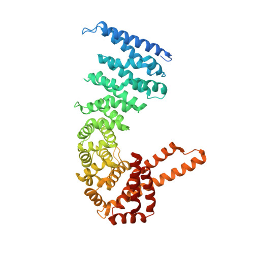
    4MFU, 4MFV

  • PubMed Abstract: 

    The hPrp19-CDC5L complex plays a crucial role during human pre-mRNA splicing by catalytic activation of the spliceosome. In order to elucidate the molecular architecture of the hPrp19-CDC5L complex, the crystal structure of CTNNBL1, one of the major components of this complex, was determined. Unlike canonical ARM-repeat proteins such as β-catenin and importin-α, CTNNBL1 was found to contain a twisted and extended ARM-repeat structure at the C-terminal domain and, more importantly, the protein formed a stable dimer. A highly negatively charged patch formed in the N-terminal ARM-repeat domain of CTNNBL1 provides a binding site for CDC5L, a binding partner of the protein in the hPrp19-CDC5L complex, and these two proteins form a complex with a stoichiometry of 2:2. These findings not only present the crystal structure of a novel ARM-repeat protein, CTNNBL1, but also provide insights into the detailed molecular architecture of the hPrp19-CDC5L complex.


  • Organizational Affiliation
    • Structural and Molecular Biology Laboratory, School of Life Sciences and Biotechnology, Kyungpook National University, Daehak-ro 80, Buk-ku, Daegu 702-701, Republic of Korea.

Macromolecule Content 

  • Total Structure Weight: 123.36 kDa 
  • Atom Count: 7,866 
  • Modeled Residue Count: 978 
  • Deposited Residue Count: 1,068 
  • Unique protein chains: 1

Macromolecules

Find similar proteins by:|  3D Structure
Entity ID: 1
MoleculeChains  Sequence LengthOrganismDetailsImage
Beta-catenin-like protein 1
A, B
534Homo sapiensMutation(s): 0 
Gene Names: C20orf33CTNNBL1PP8304
UniProt & NIH Common Fund Data Resources
Find proteins for Q8WYA6 (Homo sapiens)
Explore Q8WYA6 
Go to UniProtKB:  Q8WYA6
PHAROS:  Q8WYA6
GTEx:  ENSG00000132792 
Entity Groups
Sequence Clusters30% Identity50% Identity70% Identity90% Identity95% Identity100% Identity
UniProt GroupQ8WYA6
Sequence Annotations
Expand
Reference Sequence

Experimental Data & Validation

Experimental Data

  • Method: X-RAY DIFFRACTION
  • Resolution: 2.92 Å
  • R-Value Free:  0.305 (Depositor), 0.263 (DCC) 
  • R-Value Work:  0.233 (Depositor) 
  • R-Value Observed: 0.236 (Depositor) 
Space Group: P 31
Unit Cell:
Length ( Å )Angle ( ˚ )
a = 90.037α = 90
b = 90.037β = 90
c = 175.746γ = 120
Software Package:
Software NamePurpose
DENZOdata reduction
SCALEPACKdata scaling
MOLREPphasing
REFMACrefinement
PDB_EXTRACTdata extraction
HKL-2000data collection
HKL-2000data reduction
HKL-2000data scaling

Structure Validation

View Full Validation Report


Entry History 

Deposition Data

Revision History  (Full details and data files)

  • Version 1.0: 2014-03-12
    Type: Initial release
  • Version 1.1: 2014-04-23
    Changes: Database references
  • Version 1.2: 2017-11-15
    Changes: Refinement description
  • Version 1.3: 2023-11-08
    Changes: Data collection, Database references, Refinement description