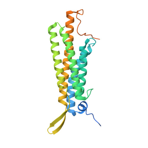
4K1O | pdb_00004k1o

Crystal structure of the alphaN-catenin actin-binding domain


Experimental Data Snapshot

  • Method: X-RAY DIFFRACTION
  • Resolution: 2.60 Å
  • R-Value Free: 
    0.231 (Depositor), 0.237 (DCC) 
  • R-Value Work: 
    0.195 (Depositor), 0.203 (DCC) 
  • R-Value Observed: 
    0.198 (Depositor) 

wwPDB Validation 3D Report Full Report

Validation slider image for 4K1O

This is version 1.2 of the entry. See complete history. 

Literature

An autoinhibited structure of alpha-catenin and its implications for vinculin recruitment to adherens junctions.

Ishiyama, N., Tanaka, N., Abe, K., Yang, Y.J., Abbas, Y.M., Umitsu, M., Nagar, B., Bueler, S.A., Rubinstein, J.L., Takeichi, M., Ikura, M.

(2013) J Biol Chem 288: 15913-15925

  • DOI: https://doi.org/10.1074/jbc.M113.453928
  • Primary Citation Related Structures: 
    4K1N, 4K1O

  • PubMed Abstract: 

    α-Catenin is an actin- and vinculin-binding protein that regulates cell-cell adhesion by interacting with cadherin adhesion receptors through β-catenin, but the mechanisms by which it anchors the cadherin-catenin complex to the actin cytoskeleton at adherens junctions remain unclear. Here we determined crystal structures of αE-catenin in the autoinhibited state and the actin-binding domain of αN-catenin. Together with the small-angle x-ray scattering analysis of full-length αN-catenin, we deduced an elongated multidomain assembly of monomeric α-catenin that structurally and functionally couples the vinculin- and actin-binding mechanisms. Cellular and biochemical studies of αE- and αN-catenins show that αE-catenin recruits vinculin to adherens junctions more effectively than αN-catenin, partly because of its higher affinity for actin filaments. We propose a molecular switch mechanism involving multistate conformational changes of α-catenin. This would be driven by actomyosin-generated tension to dynamically regulate the vinculin-assisted linkage between adherens junctions and the actin cytoskeleton.


  • Organizational Affiliation: 
    • Campbell Family Cancer Research Institute, Ontario Cancer Institute, Toronto, Ontario M5G 1L7, Canada.

Macromolecule Content 

  • Total Structure Weight: 29.39 kDa 
  • Atom Count: 1,544 
  • Modeled Residue Count: 200 
  • Deposited Residue Count: 264 
  • Unique protein chains: 1

Macromolecules

Find similar proteins by:|  3D Structure
Entity ID: 1
MoleculeChains  Sequence LengthOrganismDetailsImage
Catenin alpha-2264Mus musculusMutation(s): 0 
Gene Names: Catna2, Ctnna2
UniProt
Find proteins for Q61301 (Mus musculus)
Explore Q61301 
Go to UniProtKB:  Q61301
Entity Groups
Sequence Clusters30% Identity50% Identity70% Identity90% Identity95% Identity100% Identity
UniProt GroupQ61301
Sequence Annotations
Expand
Reference Sequence

Experimental Data & Validation

Experimental Data

  • Method: X-RAY DIFFRACTION
  • Resolution: 2.60 Å
  • R-Value Free:  0.231 (Depositor), 0.237 (DCC) 
  • R-Value Work:  0.195 (Depositor), 0.203 (DCC) 
  • R-Value Observed: 0.198 (Depositor) 
Space Group: P 32 1 2
Unit Cell:
Length ( Å )Angle ( ˚ )
a = 112.152α = 90
b = 112.152β = 90
c = 56.316γ = 120
Software Package:
Software NamePurpose
SCALEPACKdata scaling
PHENIXrefinement
PDB_EXTRACTdata extraction
HKL-3000data collection
HKL-2000data reduction
HKL-2000data scaling
PHASERphasing

Structure Validation

View Full Validation Report


Entry History 

Deposition Data

Revision History  (Full details and data files)

  • Version 1.0: 2013-05-01
    Type: Initial release
  • Version 1.1: 2013-09-04
    Changes: Database references
  • Version 1.2: 2024-02-28
    Changes: Data collection, Database references, Derived calculations