4IWQ

Crystal structure and mechanism of activation of TBK1


Experimental Data Snapshot

  • Method: X-RAY DIFFRACTION
  • Resolution: 3.00 Å
  • R-Value Free: 
    0.310 (Depositor), 0.310 (DCC) 
  • R-Value Work: 
    0.262 (Depositor), 0.260 (DCC) 
  • R-Value Observed: 
    0.264 (Depositor) 

wwPDB Validation   3D Report Full Report


Ligand Structure Quality Assessment 

Created with Raphaël 2.3.0Worse 01 BetterLigand structure goodness of fit to experimental dataBest fitted 1FVClick on this verticalbar to view details

This is version 1.2 of the entry. See complete history


Literature

Crystal structure and mechanism of activation of TANK-binding kinase 1.

Larabi, A.Devos, J.M.Ng, S.L.Nanao, M.H.Round, A.Maniatis, T.Panne, D.

(2013) Cell Rep 3: 734-746

  • DOI: https://doi.org/10.1016/j.celrep.2013.01.034
  • Primary Citation of Related Structures:  
    4IW0, 4IWO, 4IWP, 4IWQ

  • PubMed Abstract: 

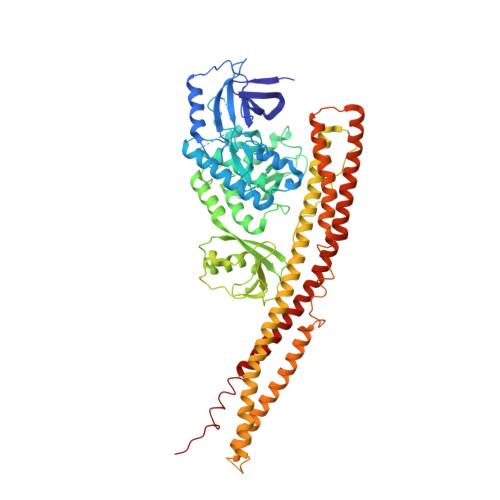
    Tank-binding kinase I (TBK1) plays a key role in the innate immune system by integrating signals from pattern-recognition receptors. Here, we report the X-ray crystal structures of inhibitor-bound inactive and active TBK1 determined to 2.6 Å and 4.0 Å resolution, respectively. The structures reveal a compact dimer made up of trimodular subunits containing an N-terminal kinase domain (KD), a ubiquitin-like domain (ULD), and an α-helical scaffold dimerization domain (SDD). Activation rearranges the KD into an active conformation while maintaining the overall dimer conformation. Low-resolution SAXS studies reveal that the missing C-terminal domain (CTD) extends away from the main body of the kinase dimer. Mutants that interfere with TBK1 dimerization show significantly reduced trans-autophosphorylation but retain the ability to bind adaptor proteins through the CTD. Our results provide detailed insights into the architecture of TBK1 and the molecular mechanism of activation.


  • Organizational Affiliation

    EMBL Grenoble, 6 Rue Jules Horowitz, Grenoble 38042, France.


Macromolecules
Find similar proteins by:  (by identity cutoff)  |  3D Structure
Entity ID: 1
MoleculeChains Sequence LengthOrganismDetailsImage
Serine/threonine-protein kinase TBK1659Homo sapiensMutation(s): 1 
Gene Names: NAKTBK1
EC: 2.7.11.1
UniProt & NIH Common Fund Data Resources
Find proteins for Q9UHD2 (Homo sapiens)
Explore Q9UHD2 
Go to UniProtKB:  Q9UHD2
PHAROS:  Q9UHD2
GTEx:  ENSG00000183735 
Entity Groups  
Sequence Clusters30% Identity50% Identity70% Identity90% Identity95% Identity100% Identity
UniProt GroupQ9UHD2
Sequence Annotations
Expand
  • Reference Sequence
Small Molecules
Ligands 1 Unique
IDChains Name / Formula / InChI Key2D Diagram3D Interactions
1FV
Query on 1FV

Download Ideal Coordinates CCD File 
B [auth A]N-{3-[(5-cyclopropyl-2-{[3-(morpholin-4-ylmethyl)phenyl]amino}pyrimidin-4-yl)amino]propyl}cyclobutanecarboxamide
C26 H36 N6 O2
UKBGBACORPRCGG-UHFFFAOYSA-N
Binding Affinity Annotations 
IDSourceBinding Affinity
1FV BindingDB:  4IWQ Kd: min: 32, max: 251 (nM) from 3 assay(s)
IC50: min: 19, max: 357 (nM) from 3 assay(s)
Experimental Data & Validation

Experimental Data

  • Method: X-RAY DIFFRACTION
  • Resolution: 3.00 Å
  • R-Value Free:  0.310 (Depositor), 0.310 (DCC) 
  • R-Value Work:  0.262 (Depositor), 0.260 (DCC) 
  • R-Value Observed: 0.264 (Depositor) 
Space Group: P 32 2 1
Unit Cell:
Length ( Å )Angle ( ˚ )
a = 135.76α = 90
b = 135.76β = 90
c = 86.5γ = 120
Software Package:
Software NamePurpose
PHENIXrefinement
PDB_EXTRACTdata extraction
ADSCdata collection
XDSdata reduction
XDSdata scaling
PHASERphasing

Structure Validation

View Full Validation Report


Ligand Structure Quality Assessment 

Created with Raphaël 2.3.0Worse 01 BetterLigand structure goodness of fit to experimental dataBest fitted 1FVClick on this verticalbar to view details

Entry History 

Deposition Data

Revision History  (Full details and data files)

  • Version 1.0: 2013-03-13
    Type: Initial release
  • Version 1.1: 2013-05-22
    Changes: Database references
  • Version 1.2: 2024-02-28
    Changes: Data collection, Database references, Derived calculations