4IVI | pdb_00004ivi

Crystal structure of a family VIII carboxylesterase.


Experimental Data Snapshot

  • Method: X-RAY DIFFRACTION
  • Resolution: 2.00 Å
  • R-Value Free: 
    0.198 (Depositor), 0.202 (DCC) 
  • R-Value Work: 
    0.182 (Depositor), 0.185 (DCC) 

wwPDB Validation 3D Report Full Report

Validation slider image for 4IVI

This is version 1.3 of the entry. See complete history

Literature

Structural basis for the beta-lactamase activity of EstU1, a family VIII carboxylesterase.

Cha, S.S.An, Y.J.Jeong, C.S.Kim, M.K.Jeon, J.H.Lee, C.M.Lee, H.S.Kang, S.G.Lee, J.H.

(2013) Proteins 81: 2045-2051

  • DOI: https://doi.org/10.1002/prot.24334
  • Primary Citation Related Structures: 
    4IVI, 4IVK

  • PubMed Abstract: 

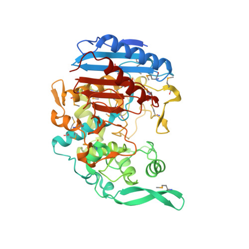
    EstU1 is a unique family VIII carboxylesterase that displays hydrolytic activity toward the amide bond of clinically used β-lactam antibiotics as well as the ester bond of p-nitrophenyl esters. EstU1 assumes a β-lactamase-like modular architecture and contains the residues Ser100, Lys103, and Tyr218, which correspond to the three catalytic residues (Ser64, Lys67, and Tyr150, respectively) of class C β-lactamases. The structure of the EstU1/cephalothin complex demonstrates that the active site of EstU1 is not ideally tailored to perform an efficient deacylation reaction during the hydrolysis of β-lactam antibiotics. This result explains the weak β-lactamase activity of EstU1 compared with class C β-lactamases. Finally, structural and sequential comparison of EstU1 with other family VIII carboxylesterases elucidates an operative molecular strategy used by family VIII carboxylesterases to extend their substrate spectrum.


  • Organizational Affiliation
    • Marine Biotechnology Research Division, Korea Institute of Ocean Science and Technology, Ansan, 426-744, Republic of Korea; Ocean Science and Technology School, Korea Maritime University, Pusan, 606-791, Republic of Korea; Department of Marine Biotechnology, University of Science and Technology, Daejeon, 305-333, Republic of Korea.

Macromolecule Content 

  • Total Structure Weight: 48.44 kDa 
  • Atom Count: 3,491 
  • Modeled Residue Count: 404 
  • Deposited Residue Count: 434 
  • Unique protein chains: 1

Macromolecules

Find similar proteins by:|  3D Structure
Entity ID: 1
MoleculeChains  Sequence LengthOrganismDetailsImage
Carboxylesterase434uncultured bacteriumMutation(s): 0 
EC: 3.1.1.1
UniProt
Find proteins for A0ACD6B8M6 (uncultured bacterium)
Explore A0ACD6B8M6 
Go to UniProtKB:  A0ACD6B8M6
Entity Groups
Sequence Clusters30% Identity50% Identity70% Identity90% Identity95% Identity100% Identity
UniProt GroupA0ACD6B8M6
Sequence Annotations
Expand
Reference Sequence

Small Molecules

Modified Residues  1 Unique
IDChains TypeFormula2D DiagramParent
MSE
Query on MSE
A
L-PEPTIDE LINKINGC5 H11 N O2 SeMET

Experimental Data & Validation

Experimental Data

  • Method: X-RAY DIFFRACTION
  • Resolution: 2.00 Å
  • R-Value Free:  0.198 (Depositor), 0.202 (DCC) 
  • R-Value Work:  0.182 (Depositor), 0.185 (DCC) 
Space Group: I 4 2 2
Unit Cell:
Length ( Å )Angle ( ˚ )
a = 149.065α = 90
b = 149.065β = 90
c = 171.131γ = 90
Software Package:
Software NamePurpose
ADSCdata collection
SOLVEphasing
CNSrefinement
HKL-2000data reduction
HKL-2000data scaling

Structure Validation

View Full Validation Report


Entry History 

Deposition Data

Revision History  (Full details and data files)

  • Version 1.0: 2013-06-19
    Type: Initial release
  • Version 1.1: 2013-10-30
    Changes: Database references
  • Version 1.2: 2018-01-24
    Changes: Structure summary
  • Version 1.3: 2024-11-20
    Changes: Data collection, Database references, Derived calculations, Structure summary