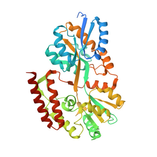
2.25 Angstrom Structure of the Extracellular Solute-binding Protein from Staphylococcus aureus in complex with Maltose.
Minasov, G., Shuvalova, L., Dubrovska, I., Winsor, J., Bagnoli, F., Falugi, F., Bottomley, M., Grandi, G., Anderson, W.F., Center for Structural Genomics of Infectious Diseases (CSGID)To be published.