4GTP | pdb_00004gtp

FTase in complex with BMS analogue 16


Experimental Data Snapshot

  • Method: X-RAY DIFFRACTION
  • Resolution: 2.75 Å
  • R-Value Free: 
    0.226 (Depositor), 0.222 (DCC) 
  • R-Value Work: 
    0.177 (Depositor) 
  • R-Value Observed: 
    0.179 (Depositor) 

Starting Model: experimental
View more details

wwPDB Validation 3D Report Full Report

Validation slider image for 4GTP

Ligand Structure Quality Assessment 


This is version 1.1 of the entry. See complete history

Literature

Development of Selective, Potent RabGGTase Inhibitors

Stigter, E.A.Guo, Z.Bon, R.S.Wu, Y.W.Choidas, A.Wolf, A.Menninger, S.Waldmann, H.Blankenfeldt, W.Goody, R.S.

(2012) J Med Chem 55: 8330-8340

  • DOI: https://doi.org/10.1021/jm300624s
  • Primary Citation Related Structures: 
    4GTM, 4GTO, 4GTP, 4GTQ, 4GTR, 4GTS, 4GTT, 4GTV

  • PubMed Abstract: 

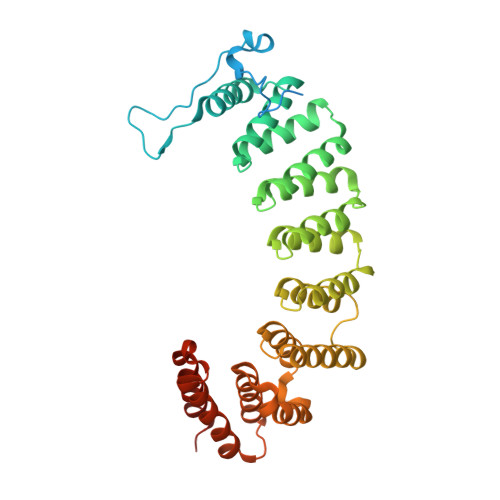
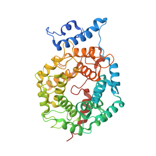
    Members of the Ras superfamily of small GTPases are frequently mutated in cancer. Therefore, inhibitors have been developed to address the acitivity of these GTPases by inhibiting their prenylating enzymes FTase, GGTase I, and RabGGTase. In contrast to FTase and GGTase I, only a handful of RabGGTase inhibitors have been developed. The most active RabGGTase inhibitor known until recently was an FTase inhibitor which hit RabGGTase as an off-target. We recently reported our efforts to tune the selectivity of these inhibitors toward RabGGTase. Here we describe an extended set of selective inhibitors. The requirements for selective RabGGTase inhibitors are described in detail, guided by multiple crystal structures. In order to relate in vitro and cellular activity, a high-throughput assay system to detect the attachment of [(3)H]geranylgeranyl groups to Rab was used. Selective RabGGTase inhibition allows the establishment of novel drug discovery programs aimed at the development of anticancer therapeutics.


  • Organizational Affiliation
    • Department of Chemical Biology, Max Planck Institute of Molecular Physiology, Otto-Hahn-Strasse 11, 44227 Dortmund, Germany.

Macromolecule Content 

  • Total Structure Weight: 93.22 kDa 
  • Atom Count: 6,072 
  • Modeled Residue Count: 718 
  • Deposited Residue Count: 804 
  • Unique protein chains: 2

Macromolecules

Find similar proteins by:|  3D Structure
Entity ID: 1
MoleculeChains  Sequence LengthOrganismDetailsImage
Protein farnesyltransferase/geranylgeranyltransferase type-1 subunit alpha377Rattus norvegicusMutation(s): 0 
Gene Names: Fnta
EC: 2.5.1.58 (PDB Primary Data), 2.5.1.59 (PDB Primary Data)
UniProt
Find proteins for Q04631 (Rattus norvegicus)
Explore Q04631 
Go to UniProtKB:  Q04631
Entity Groups
Sequence Clusters30% Identity50% Identity70% Identity90% Identity95% Identity100% Identity
UniProt GroupQ04631
Sequence Annotations
Expand
Reference Sequence
Find similar proteins by:|  3D Structure
Entity ID: 2
MoleculeChains  Sequence LengthOrganismDetailsImage
Protein farnesyltransferase subunit beta427Rattus norvegicusMutation(s): 0 
Gene Names: Fntb
EC: 2.5.1.58
UniProt
Find proteins for Q02293 (Rattus norvegicus)
Explore Q02293 
Go to UniProtKB:  Q02293
Entity Groups
Sequence Clusters30% Identity50% Identity70% Identity90% Identity95% Identity100% Identity
UniProt GroupQ02293
Sequence Annotations
Expand
Reference Sequence

Small Molecules

Ligands 4 Unique
IDChains Name / Formula / InChI Key2D Diagram3D Interactions
7TP

Query on 7TP


Download:Ideal Coordinates CCD File
F [auth B]5-{(3R)-3-(4-hydroxybenzyl)-4-[(4-methoxyphenyl)sulfonyl]-1-[(1-methyl-1H-imidazol-5-yl)methyl]-2,3,4,5-tetrahydro-1H-1,4-benzodiazepin-7-yl}furan-2-carbaldehyde
C33 H32 N4 O6 S
HXGORCFYRJWSGR-AREMUKBSSA-N
FPP

Query on FPP


Download:Ideal Coordinates CCD File
E [auth B]FARNESYL DIPHOSPHATE
C15 H28 O7 P2
VWFJDQUYCIWHTN-YFVJMOTDSA-N
DMS

Query on DMS


Download:Ideal Coordinates CCD File
C [auth A],
G [auth B],
H [auth B],
I [auth B]
DIMETHYL SULFOXIDE
C2 H6 O S
IAZDPXIOMUYVGZ-UHFFFAOYSA-N
ZN

Query on ZN


Download:Ideal Coordinates CCD File
D [auth B]ZINC ION
Zn
PTFCDOFLOPIGGS-UHFFFAOYSA-N

Experimental Data & Validation

Experimental Data

  • Method: X-RAY DIFFRACTION
  • Resolution: 2.75 Å
  • R-Value Free:  0.226 (Depositor), 0.222 (DCC) 
  • R-Value Work:  0.177 (Depositor) 
  • R-Value Observed: 0.179 (Depositor) 
Space Group: P 61
Unit Cell:
Length ( Å )Angle ( ˚ )
a = 171.65α = 90
b = 171.65β = 90
c = 70.005γ = 120
Software Package:
Software NamePurpose
MOSFLMdata reduction
PHASESphasing
REFMACrefinement
XDSdata reduction
XSCALEdata scaling

Structure Validation

View Full Validation Report


Ligand Structure Quality Assessment 


Entry History 

Deposition Data

Revision History  (Full details and data files)

  • Version 1.0: 2012-10-24
    Type: Initial release
  • Version 1.1: 2023-11-08
    Changes: Data collection, Database references, Derived calculations, Refinement description